 |
|
Project 2: Hygiene Hypothesis
Each year in this
country more than 13,000 young people are diagnosed with type 1
diabetes (T1D).
In this autoimmune disease, insulin-producing b
cells are destroyed by CD4 and CD8 T cells infiltrating
the pancreatic islets, causing defects in blood glucose homeostasis and
ultimately vascular and neurological complications. T1D is potentially
life
threatening, and an increasingly significant public health problem
worldwide,
particularly in western European countries where there is an alarming
decrease
in the age of onset. The “hygiene hypothesis” links the increase in
autoimmune
phenomena in humans to excessively sanitary conditions early in life.
In
retrospective studies that seek to link the relationship of typical
childhood
infectious diseases to T1D, exposure to Streptococcus
pyogenes
(GAS) had a significant negative correlation with
acquisition of T1D. In
rodent
models treatment with GAS preparations leads to diabetes resistance. In
GAS
infections antibodies are made against cell wall-associated
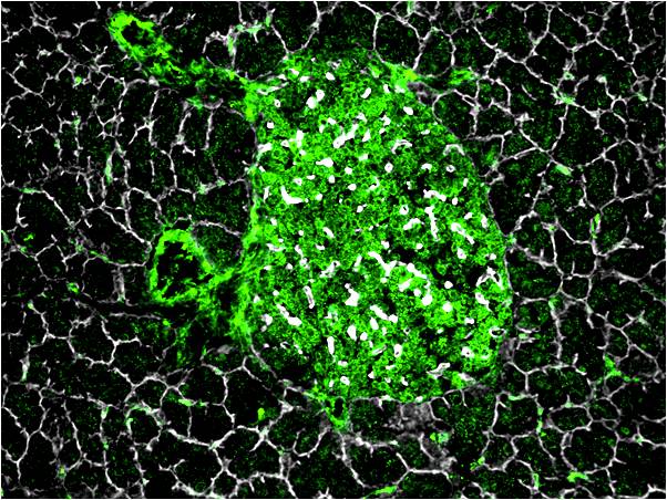
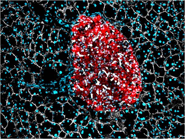
N-acetyl-D-glucosamine (GlcNAc). We will test the hypothesis that
alternative GlcNAc-specific
B lymphocyte activities can influence the development of autoimmune
diabetes by
contributing to protection or acceleration of disease. Anti-GlcNAc antibodies
also bind proteins and insulin-containing secretory granules
enriched in pancreatic β cells. We
will examine a role for GlcNAc specific B lymphocytes in modulating T1D
in
mouse models and test the hypothesis that anti-GlcNAc lymphocytes
protect
against T1D by generating anti-GlcNAc antibodies that dampen the
autoimmune
response to GlcNAcylated molecules associated with β islet cells. In an
alternative context,
we propose that anti-GlcNAc B-lymphocytes can be involved on presenting
GlcNAcylated β cell autoantigens to diabetogenic
T cells. In
Aim 1 we will determine how early immunization and timed passive
anti-GlcNAc
antibody influence the rate and severity of T1D progression in NOD
mice. Aim 2
will study the effects of antibodies on antigen-presenting cell
activation of
diabetogenic T cells in vitro. Finally in Aim 3 we will determine the
mechanisms
of protection against T1D development in intact NOD mice. |
|
|
 |
| Relevance The knowledge of how a common infectious organism, GAS, alters an individual’s propensity to develop type I diabetes (T1D) will be applied to understanding mechanisms involved in T1D induction and progression in the NOD model which simulates many aspects of human T1D. The long-term goal of this project is to identify factors that can influence the progression of T1D in susceptible individuals, which may improve T1D diagnosis and development of immunization strategies for prevention of T1D. |
(B) Characterization of Pancreas-Infiltrating GlcNAc-Reactive B cell Clonotypes in Human T1D Very little is known regarding the specificity and clonal heterogeneity of islet infiltrating B cells in human T1D, and the progression of successive targets within the B cell response of T1D remains unknown In addition to the studes of our mouse models we plan to develop reagents that enable longitudinal tracking of clonal frequencies represented in GlcNAc-reactive B cell pool of pre-autoantibody positive T1D patients. Many innate-like B cell antigen specificities, such as GlcNAc, exhibit high levels of clonal restriction in the human population. In cases of these B cell clonotypes, anti-idiotypic antibodies may be used to identify B cell receptor-restricted epitopes that correlate with certain clonal and fine-specificity antibody qualities. The goal of this aim is to translate our findings in mice regarding T1D-suppressive GlcNAc-reactive B cells into diagnostic tools through the generation of anti-idiotypic antibody reagents specific for human GlcNAc-reactive B cells. Following our findings in NOD mice, we propose that GlcNAc-reactive B cells clones will be recoverable from pancreatic tissue of T1D patients. The goal of this aim is to pair our antibody cloning technology, with Laser Microdissection Microscopy to identify GlcNAc-reactive B cell clonotypes recovered from human pancreas biopsies. Identification of cryptic glycan-reactive B cell clonotypes, particularly class-switched clonotypes involved in T1D progression, will facilitate the development and validation of specific anti-Id reagents to monitor the activation and differentiation of disease-associated clonotypes. |