 19 yo F w/ fam. h/o cardiomyopathy, arrhythmia & renal dz. Cr 0.8, BUN 11. UA neg. Renal bx.
What's the underlying condition?
A. Alport syndrome
B. Fabry disease
C. Lipoprotein glomerulopathy
D. Lecithin-cholesterol acyltransferase deficiency
Answer
B: Fabry disease
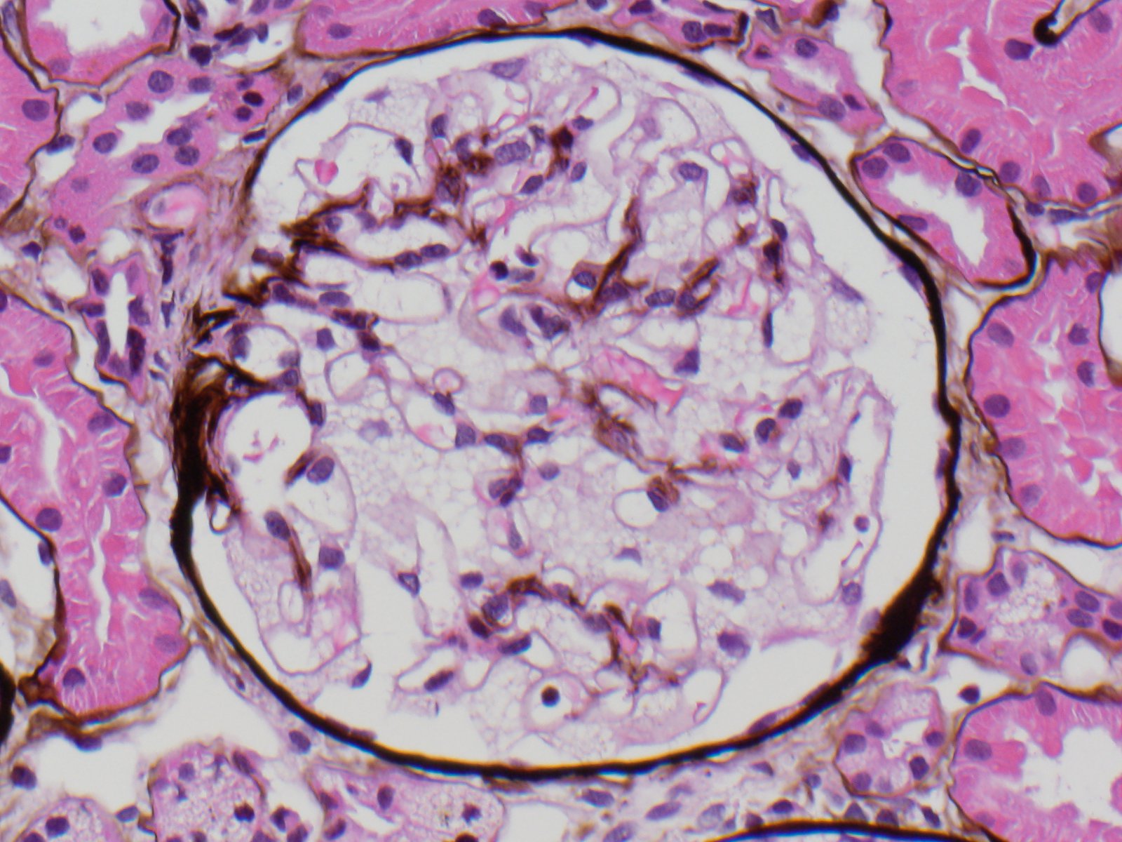
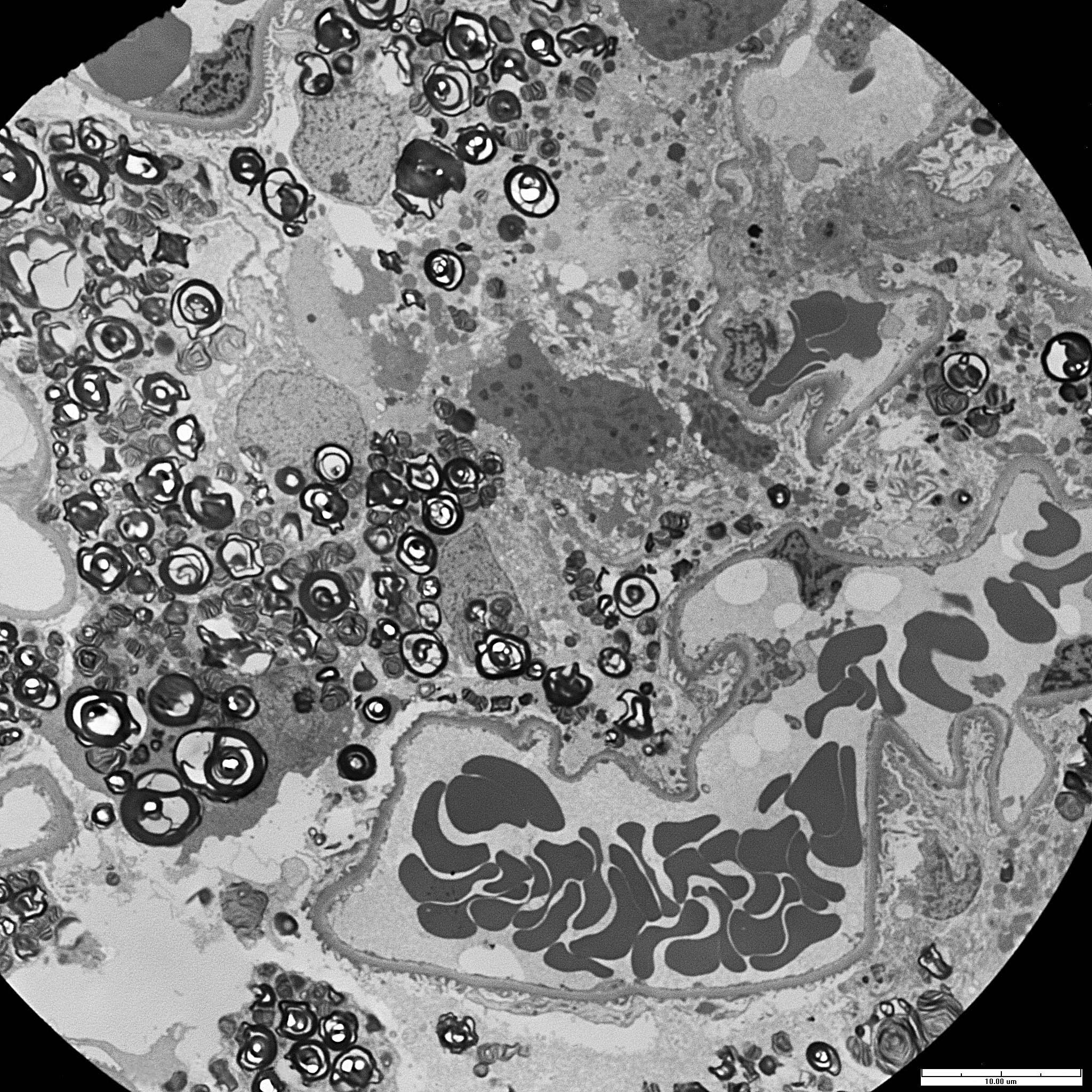
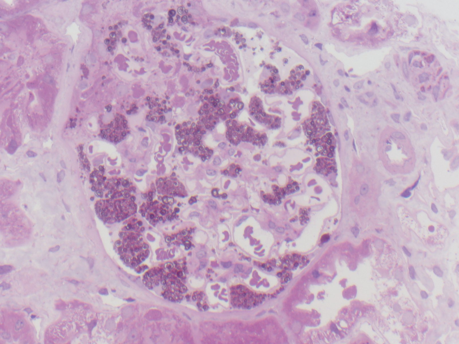
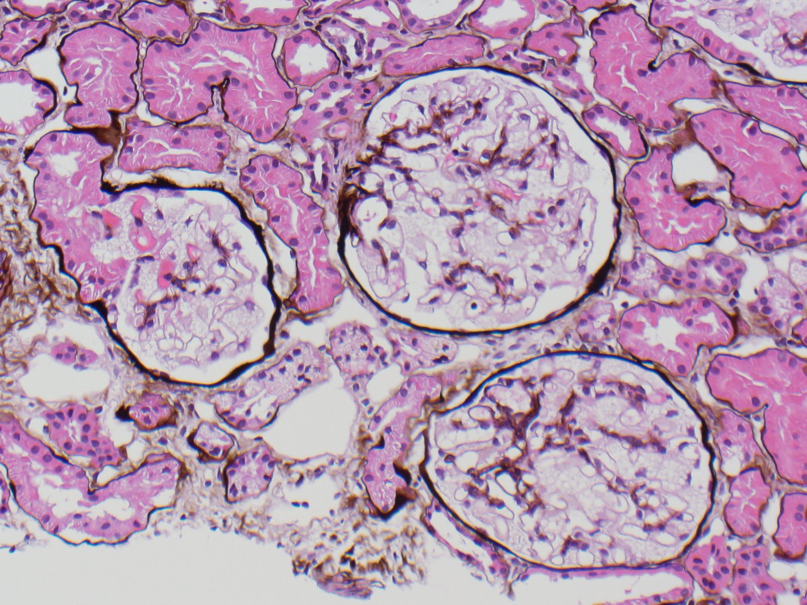
Fabry disease is an X-linked deficiency of alpha-galactosidase A leading to intracellular accumulation of glycosphingolipids. Clinical manifestations are systemic because this enzyme is present in lysosomes throughout the body; they include painful neuropathies, skin lesions (classically angiokeratomas), cardiovascular and renal disease (commonly proteinuria). By light microscopy, the accumulated galactosyl ceramide appears as vacuolization of podocytes and tubular cells because it is extracted by the xylene used in processing. By electron microscopy, lamellated inclusions called myelin or zebra bodies are most prominently present in podocytes, but are also variably present in endothelium, tubules and interstitial cells. Genetically engineered alpha-galactosidase A can now be used to treat patients, resulting in decreased disease manifestation.
Classical Alport syndrome is an X-linked dominant glomerular basement membrane disorder due to a mutation of alpha 5 type collagen and usually presents as hematuria. The key diagnostic feature is a thin or basket-weave appearance of the glomerular basement membrane by electron microscopy. Lipoprotein glomerulopathy present with proteinuria and more commonly in Japanese or Chinese males. The characteristic microscopic lesion is of intracapillary lipoprotein thrombi distending and enlarging glomeruli, and by electron microscopy there are vacuolated or granular deposits and thrombi. Lecithin-cholesterol acyltransferase deficiency is autosomal recessive and patients present with proteinuria, hyperlipidemia, corneal opacities and atherosclerosis. By light microscopy the capillary walls are thickened, the basement membranes have an irregular bubbly appearance and foam cells can be present in the mesangial and capillary areas. Electron microscopy reveals lacunae in basement membranes and striated dense membranous structures in the mesangium.
References
Fogo, Agnes B., and Michael Kashgarian. "1." Diagnostic Atlas of Renal Pathology. Philadelphia, PA: Elsevier, 2011. 177-188, 273-283. Print.
Contributed by: Frida Rosenblum, M.D.