Case History
22-year-old man with visual field deficit. Suprasellar mass resected which showed nuclear expression of beta-catenin.
- Anaplastic meningioma, WHO grade III
- Adamantinomatous craniopharyngioma
- Erosive basosquamous carcinoma
- Pilomatrix carcinoma
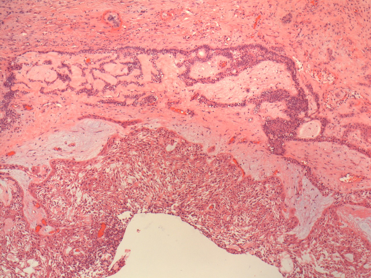
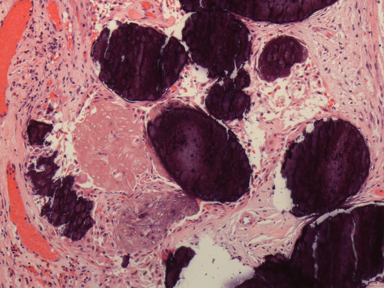
Answer: B: Adamantinomatous craniopharyngioma
Adamantinomatous craniopharyngioma, a WHO grade I neoplasm arising from Rathke pouch remnants, typically occurs in the suprasellar cistern near the base of brain and has cystic components, wet keratin, and numerous microcalcifications. Nuclear beta-catenin staining is a surrogate biomarker for the CTNNB1 gene mutation that occurs in adamantinomatous craniopharyngioma.