Case History
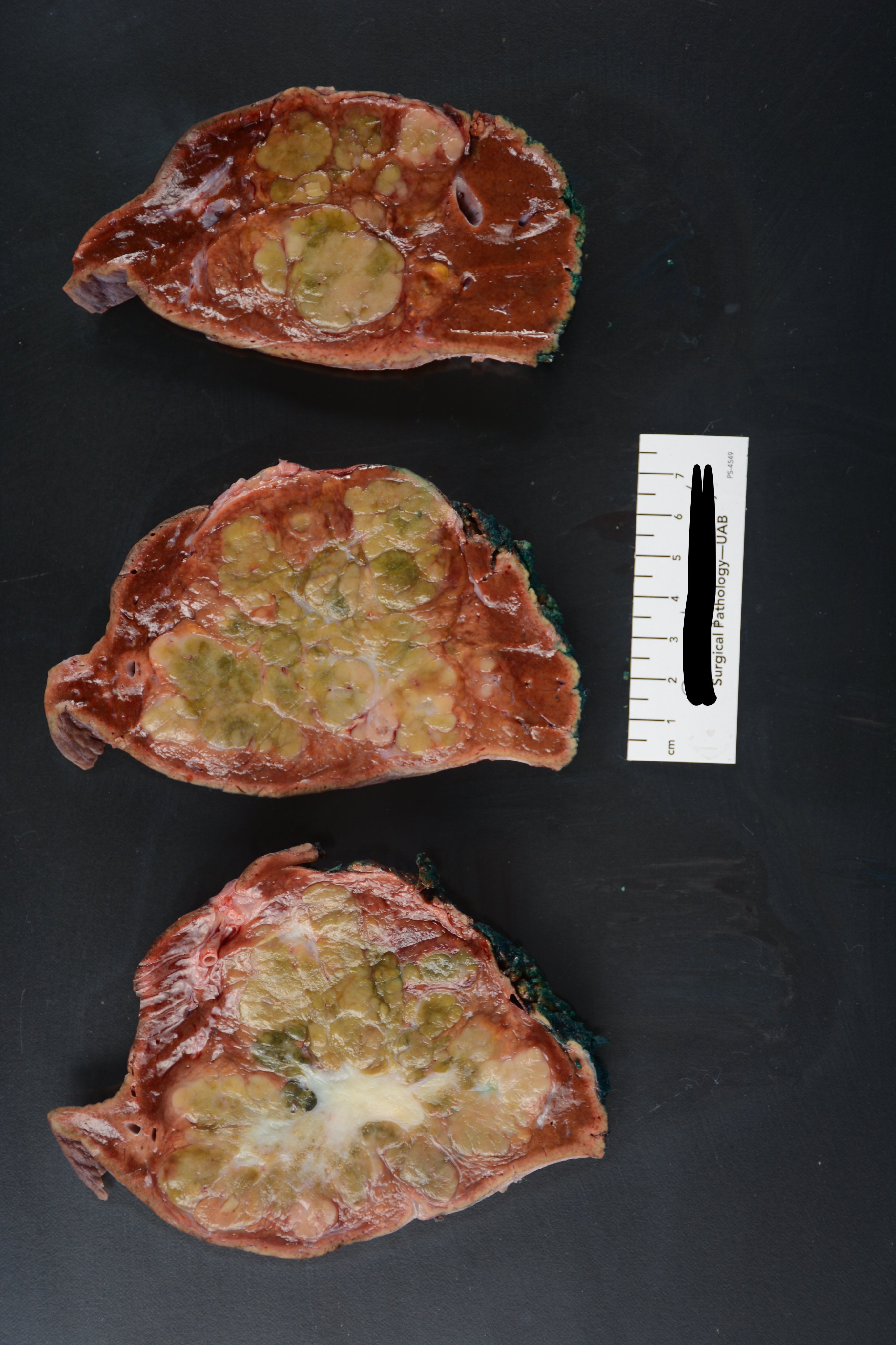
27-year-old female with incidentally discovered 9.4-cm left lobe liver mass on MRI. There is no underlying liver disease, and the background liver is non-cirrhotic. Serum AFP is normal. Hepatitis panel is non-reactive.
What is your best diagnosis?
- Focal nodular hyperplasia
- Fibrolamellar hepatol cl ca
- Hepatolcellular ca
- Cholangiocarcinoma
Answer: B. Fibrolamellar hepatocellular carcinoma (FL-HCC)
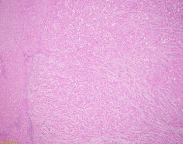
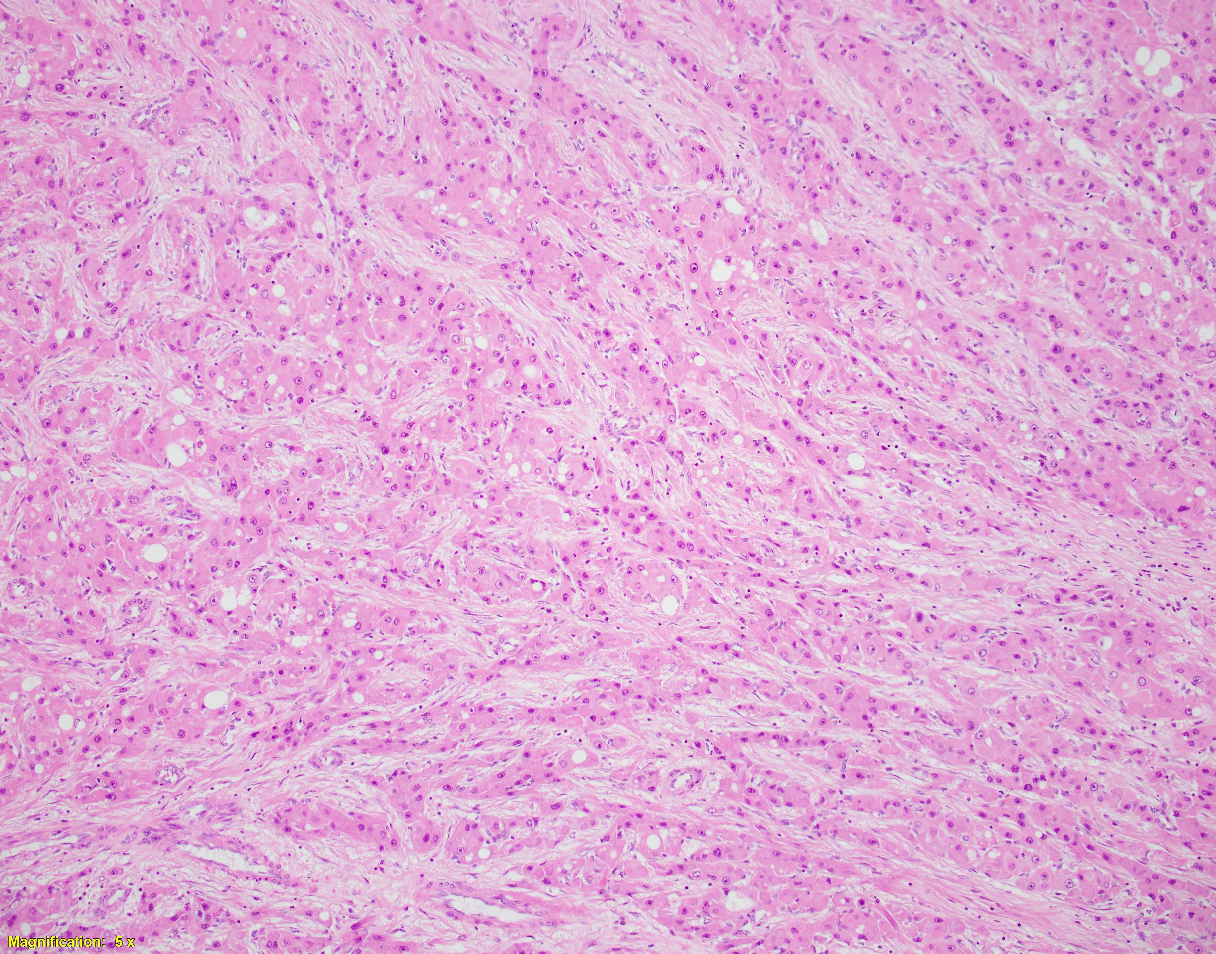
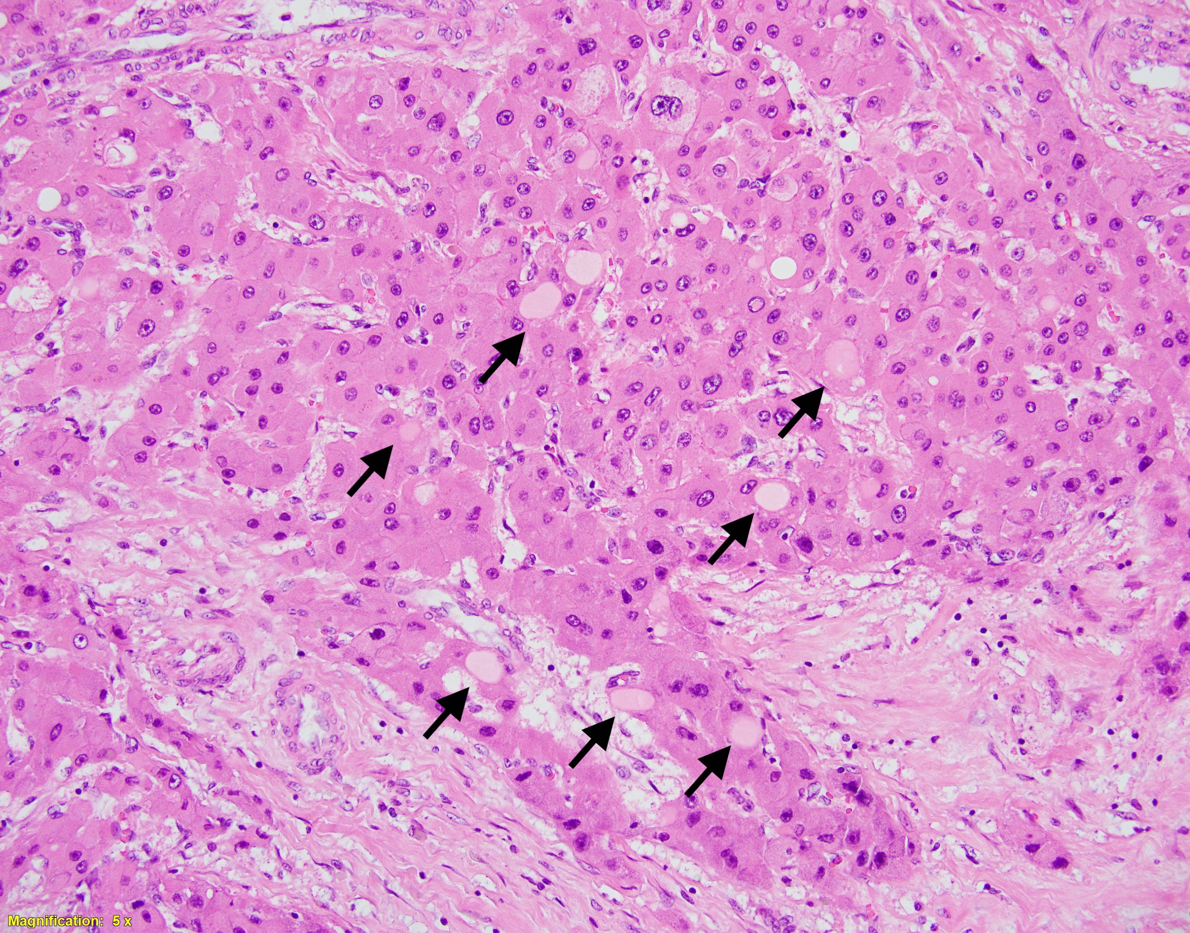
The correct answer is B. Fibrolamellar hepatocellular carcinoma (FL-HCC) is a distinct subtype of hepatocellular carcinoma. The molecular underpinning involves a DNAJB1-PRKACA translocation that creates a fusion (onco-)gene leading to constitutive activation of protein kinase A. A FISH assay can detect this (see reference 1). FL-HCC classically occurs in a young adult, typically under 50 years of age, without background liver disease. Serum AFP is classically normal range. Histologically, the tumor shows prominent dense fibrosis and parallel rays (-“lamellae”) of tumor cells along the collagen bands. The tumor cells are polygonal, contain abundant eosinophilic granular cytoplasm, vesicular chromatin, and prominent nucleoli. Pale bodies in the cytoplasm (notated by arrows) are commonly seen in FL-HCC but are not specific for this entity. Focal nodular hyperplasia (FNH) is also in the differential given that a central scar can be seen in both FL-HCC and FNH. Classic features of FNH such as bile ductular proliferation with inflammatory infiltrates at septa/periphery of the tumor are not seen. A reticulin stain (not shown) can help exclude an adenoma, as reticulin will be retained in an adenoma vs. fragmented and thickened plates (generally >3 hepatocytes thick) in HCC. Cholangiocarcinoma is incorrect answer due to absence of supporting histologic features such as gland formation and desmoplastic stroma.
References:
- Graham RP, Yeh MM, Lam-Himlin D, et al. Molecular testing for the clinical diagnosis of fibrolamellar carcinoma. Mod Pathol. 2018;31(1):141–149. doi:10.1038/modpathol.2017.103
- WHO Classification of Tumours 5th Edition: Digestive System Tumours.
International Agency for Research on Cancer 2019. pgs 229-239.