Case History
A 30-year-old man presented with a left elbow mass. A biopsy showed a small, blue, round cell tumor. The lesional cells were positive for myoD1, with very limited expression of desmin and myogenin.
What is the diagnosis?
- Extraskeletal osteosarcoma
- Ewing sarcoma
- Embryonal rhabdomyosarcoma
- Sclerosing rhabdomyosarcoma
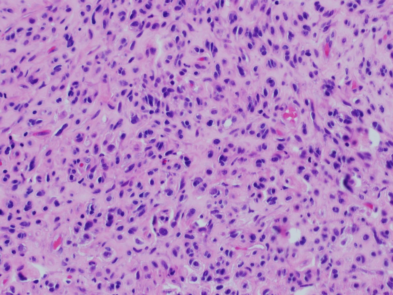
The answer is “D”, Sclerosing rhabdomyosarcoma
Spindle cell/sclerosing rhabdomyosarcoma (RMS) is uncommon. It more frequently affects male, with a male to female ratio of up to 6:1. The common locations for spindle cell RMS are paratesticular region in children and head/neck region in adults, whereas sclerosing morphology in both age groups more often involves the limbs, although other anatomic sites can rarely be affected, such as viscera and retroperitoneum. Sclerosing RMS may show very limited expression of desmin and myogenin, but is often strongly positive for myoD1. Recurrent myoD1 mutations are commonly seen in sclerosing RMS and are associated with worse prognosis. PAX3- and PAX7-FOXO1 fusions are virtually always absent.
Embryonal RMS is the most common subtype of RMS, more frequently affect Caucasians (80%). It is mostly seen in patients aged <15 years. The most common locations are head/neck region (~50%) and genitourinary system (~50%), and rarely it may occur in extremities (<9%). Histologically, embryonal RMS shows various stages of myogenesis, with variable rhabdomyoblasts. PAX3- and PAX7-FOXO1 fusions characteristics of alveolar RMS are always absent.
Ewing sarcomas are also small, blue, round cell tumors thus histologically may mimic RMS. It is driven by pathognomonic and etiological TEF-ETS gene fusions, the most common alteration being EWSR1-FLI1 translocation.
The defining feature of osteosarcoma is the production, albeit sometimes subtle, of osteoid/bone matrix by tumor cells.
References
- Fletcher CDM, Bridge, JA, Hogendoorn PCW, Mertens F. WHO Classification of Tumors of Soft Tissue and Bone. Lyon, France: IARC Press; 2013.
- Agaram NP, et al. Recurrent MYOD1 Mutations in Pediatric and Adult Sclerosing and Spindle Cell Rhabdomyosarcomas: Evidence for a Common Pathogenesis. Genes Chromosomes Cancer. 2014 Sep;53(9):779-87.
Case contributed by: Shi Wei, M.D., Ph.D., Professor, Anatomic Pathology