Case History
This is a 41 year old man with admitted from an outside hospital for encephalopathy. Chest x-ray shows multiple nodules suggestive of disseminated tuberculosis.
What is the diagnosis?
- Tuberculosis
- Histoplasmosis
- Coccidomycosis
- Blastomycosis
The answer is “C”,Coccidiomycosis (Example “A”, Granular cell tumor)
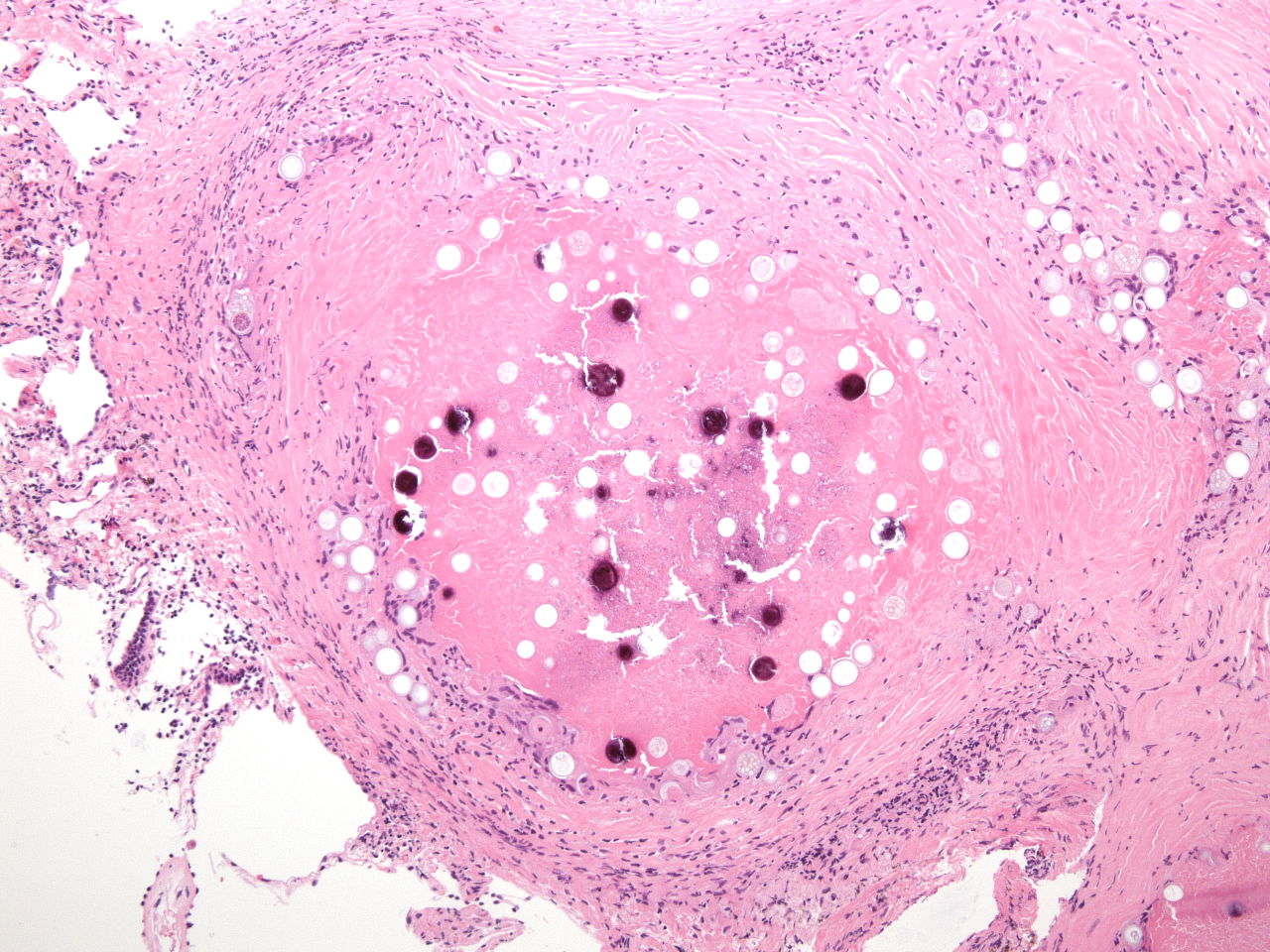
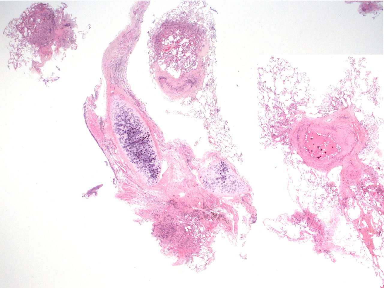
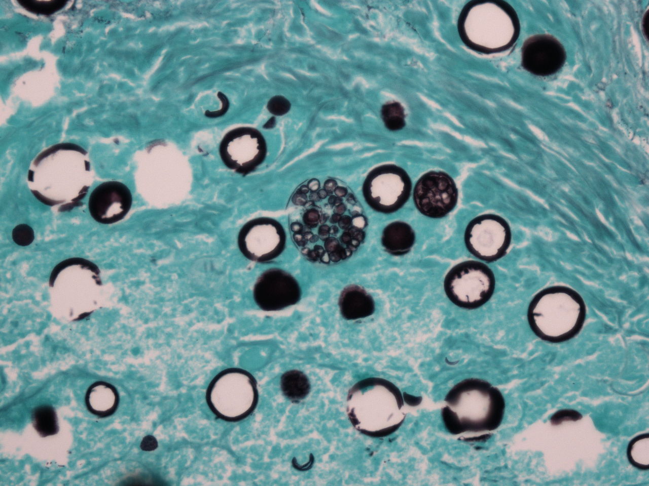
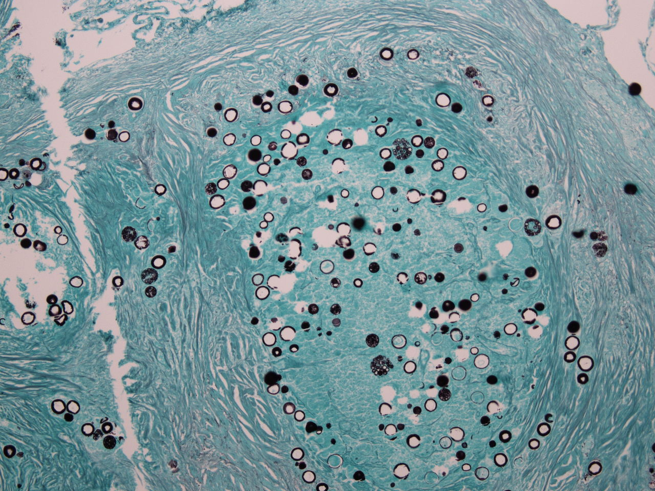
The H&E image shows fragments of peribronchial and alveolar tissue. A prominent granuloma with internal yeast and calcified yeast is seen in the largest alveolar fragment. A few of the spaces appear to have internal structure. This is seen best on the silver stain. At high magnificationof the silver stain the internal structure can be identified as daughter cells making the diagnosis of Coccidiomycosis. The patient’s history was later found to include residence in Mexico.
References
Guarner J, Brandt ME. Histopathologic diagnosis of fungal infections in the 21st century. Clin Microbiol Rev. 2011;24(2):247‐280. doi:10.1128/CMR.00053-10
Case contributed by: Thomas Winokur, M.D., Ph.D., Professor, Anatomic Pathology