Case History
A 50-year old male presents with a unilocular radiolucent lesion in the left anterior mandible noted on dental x-ray. A biopsy is performed and representative sections are shown.
What is the diagnosis?
- Mucoepidermoid carcinoma
- Periapical/radicular cyst
- Odontogenic keratocyst
- Glandular odontogenic cyst
Answer: D. Glandular odontogenic cyst
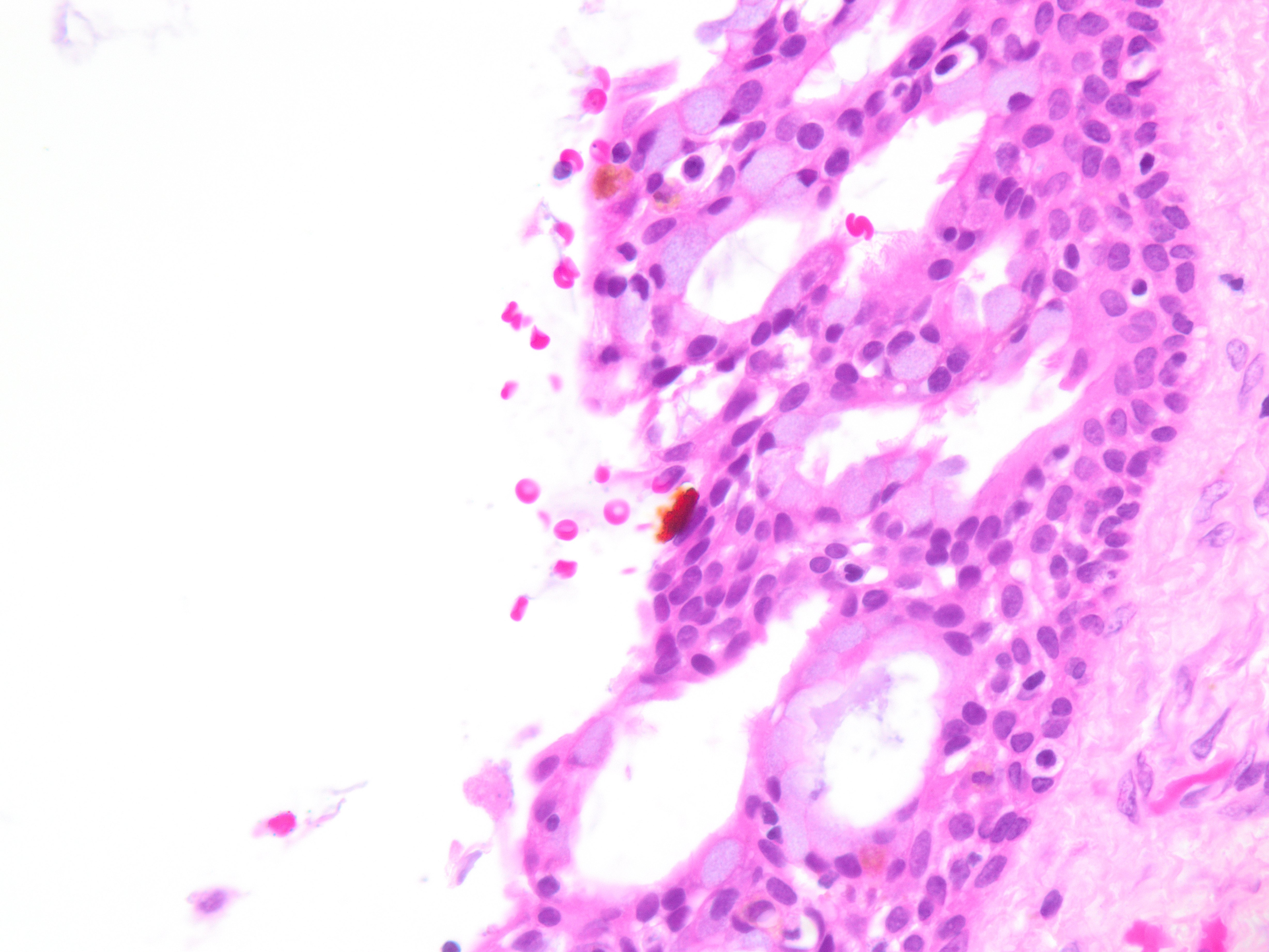
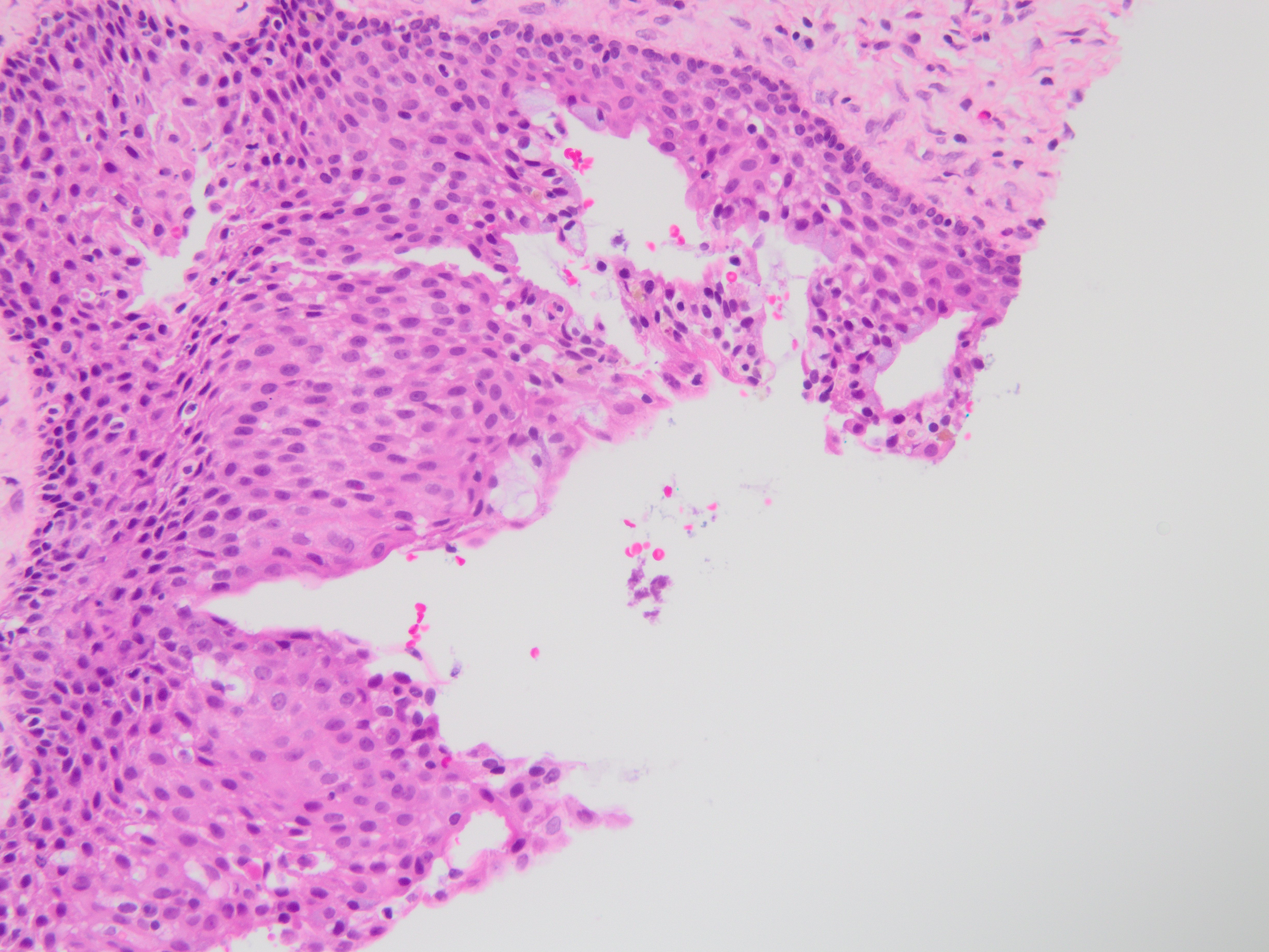
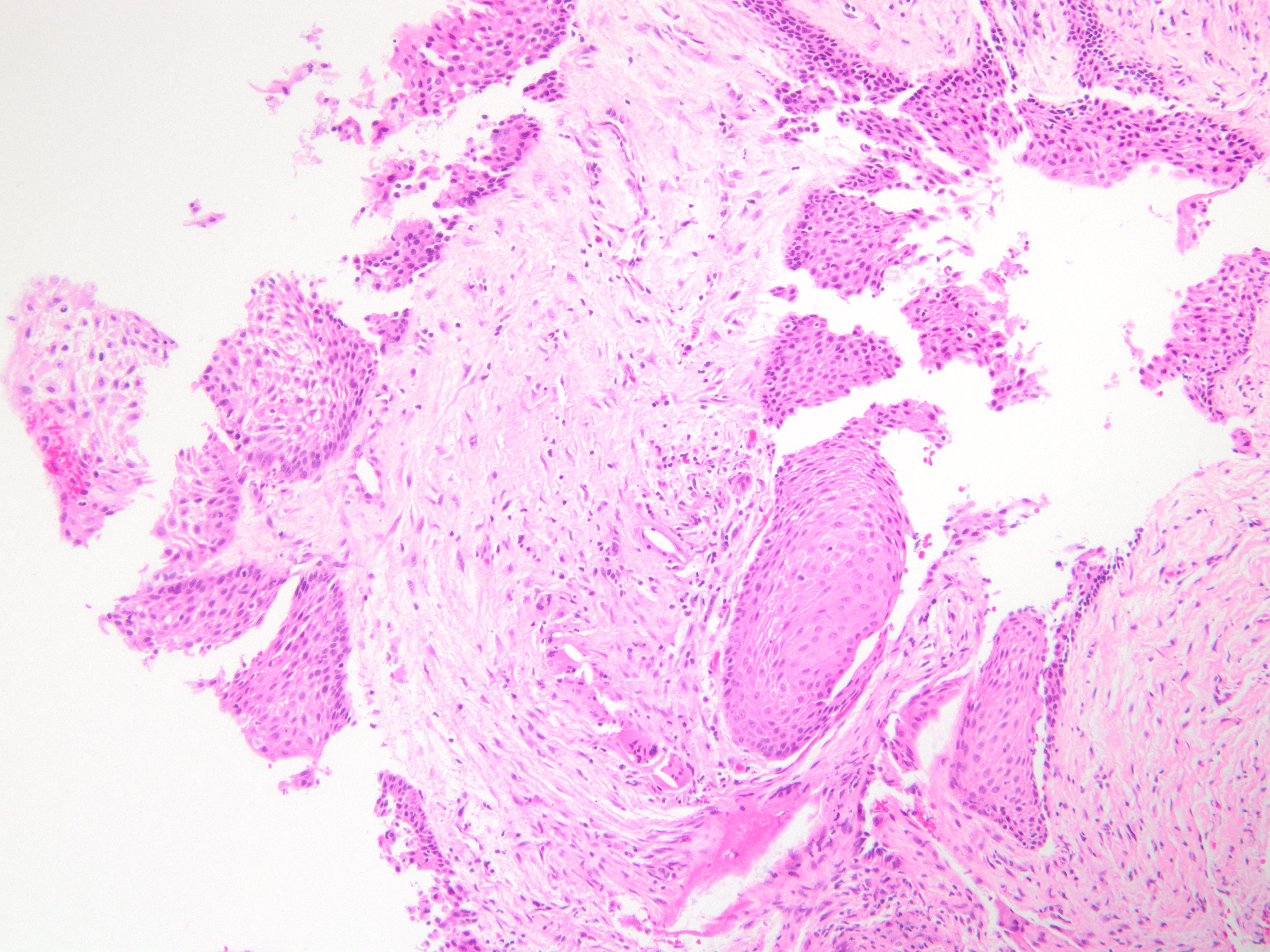
Brief explanation of the answer: Sections show cyst lining of variable thickness with mucous cells, ciliated cells, eosinophilic cuboidal cells, and clear cells. There are multiple compartments. Microcysts, papillary projections and epithelial spheres are present. These features are consistent with glandular odontogenic cyst. Resection is recommended due to high recurrence rate.
No basal cell palisading or parakeratin is noted, excluding odontogenic keratocyst. Periapical and radicular cysts tend to have a squamous lining with abundant inflammation. Mucoepidermoid carcimoma tends to be more proliferative with invasive features, and also shows intermediate and epidermoid cells.