Case History
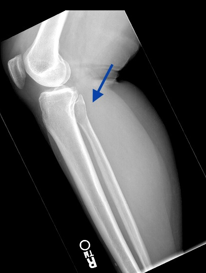
A 56-year-old woman presented complaining of right lateral knee pain that is aggravated by driving. A plain film of the right knee is provided. This is a biopsy of a fibula mass.
What is the diagnosis?
A. Brown tumor
B. Aneurysmal bone cyst
C. Giant cell tumor of bone
D. Giant cell-rich osteosarcoma
Answer: C. Giant cell tumor of bone
Discussion:
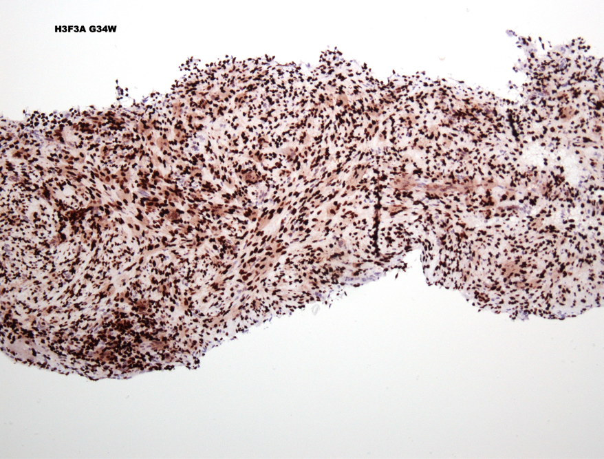
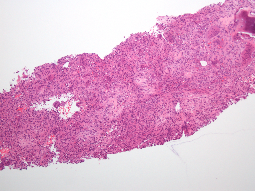
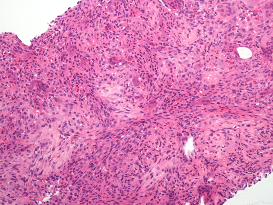
Sections show a tumor with numerous giant cells and a mononuclear component. The histologic differential diagnosis includes all of the listed lesions. Brown tumor and giant cell tumor are histologically indistinguishable. Aneurysmal bone cyst should show blood filled spaces and osteosarcoma should show tumor osteoid, however a needle biopsy can miss those features. The radiograph shows a well demarcated lesion that involves the epiphysis but extends to the metaphysis. The radiographic differential based on the plain film and CT favored a round cell tumor or metastasis. Fortunately this diagnosis can be aided by immunohistochemistry. Antibody H3.3 G34W highlights cells with the histone mutation characteristic of giant cell tumors of bone that occur in the axial skeleton. Other mutations can be seen in tumors of small bones.
WHO Classification of Tumors Editorial Board. Soft tissue and bone tumors. 2020, 440-445
Amary F, Berisha F, Ye H, Gupta M, Gutteridge A, Baumhoer D, Gibbons R, Tirabosco R, O'Donnell P, Flanagan AM. H3F3A (Histone 3.3) G34W Immunohistochemistry: A Reliable Marker Defining Benign and Malignant Giant Cell Tumor of Bone. Am J Surg Pathol. 2017 Aug;41(8):1059-1068. doi: 10.1097/PAS.0000000000000859. PMID: 28505000; PMCID: PMC5510691.