
Nanodiamonds designed to toughen artificial joints also might prevent the inflammation caused when hardworking metal joints shed debris into the body, according to an early study published this week in the journal Acta Biomaterialia.
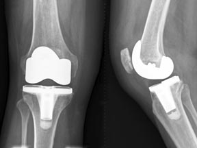
In the race to create longer-lasting and less-painful artificial joints, University of Alabama at Birmingham researchers are exploring whether nanodiamond coatings can reduce wear on joints made of metal alloys. The work is important because, according to the American Academy of Orthopedic Surgeons, more than 418,000 knee replacements and 328,000 hip replacements are performed in the United States each year; the numbers are expected to balloon as the nation’s population ages.
Joint wear generates debris that can cause pain, limit mobility and hasten joint failure. Debris particles from metal surfaces are absorbed by scavenging immune cells called macrophages, which then secrete chemicals that cause swelling and pain. This inflammation turns on bone-eating cells near implants, and bone-loss increases the likelihood implants will break loose and require a second surgery.
Diamond coatings may end the shedding of metal debris, but the constant grinding force within joints can cause even nanodiamonds to shed some particles. Past studies suggest that diamonds shed less debris and smaller particles; but, with applications emerging in drug delivery and bio-imaging, the consequences of particle build-up in organs needs to be known.
Based on the way nanodiamonds interact with macrophages in a dish, the study authors suggest that the usual size and concentration of wear debris should cause neither inflammation nor toxicity. The macrophages that engulf smaller nanodiamonds release fewer inflammatory chemicals than those encountering larger particles shed by the metal and polymer surfaces of conventional implants.
“Our results add to the early evidence that nanodiamonds are indeed nontoxic in living cells,” said Vinoy Thomas, Ph.D., research associate in the Department of Physics within the UAB College of Arts & Sciences, and corresponding author of the study. “The next step will be to conduct experiments to confirm where nanodiamond particles of varying sizes and concentrations end up, and if buildup at those destinations is safe.”
The authors cited a previous study in mice that revealed 60 percent of injected nanodiamond particles are deposited in the liver within a half hour of dosing; the remainder was deposited in the spleen and lungs. Those exploring nanodiamonds as delivery vehicles for drugs would be counting on these tendencies.
Fewer the better
In the current study, macrophages were exposed to synthetic nanodiamonds of varying sizes (6, 60, 100, 250 and 500 nanometers) and concentrations (0, 10, 50, 100, 200 micrograms per milliliter).
At concentrations of less than 50 micrograms per milliliter of solution (at which they typically occur as debris), nanodiamonds were not toxic to macrophages, which continued to thrive and metabolize energy regardless of particle size, according to the study authors. Once the concentration of nanodiamonds exceeded 200 micrograms per milliliter, macrophage viability dropped by up to 50 percent regardless of particle size.
In addition, nanodiamond exposure significantly reduced expression of several genes known to play roles in inflammation and related bone loss when compared to metal and plastic particles, including tumor necrosis factor (TNF)-?, interleukin (IL)-1?, chemokine Ccl2 and platelet derived growth factor (PDGF). Researchers contend that smaller size and lower concentration meant nanodiamonds were engulfed by macrophages that released fewer inflammatory chemicals that turned on fewer harmful genes.
“Past studies on diamond-joint surfaces have shown a marked reduction in wear-debris volume compared to first-generation alloy and polyethylene joint parts, but the work continues to ensure they are safe,” said Yogesh Vohra, Ph.D., director of the UAB Center for Nanoscale Materials and Biointegration and senior author on the study. “We hope the reduced wear volume and particle size expected for diamond articulation will represent a major advance over conventional orthopedic bearings.”
“This study provides the insight necessary for us to continue our nano-toxicological evaluation of nanodiamond particles,” said Namasivayam Ambalavanan, M.D., professor in the Department of Pediatrics within the UAB School of Medicine, and study co-author.
The work was funded by the National Institute of Arthritis and Musculoskeletal and Skin Diseases, part of the National Institutes of Health. Also making important contributions as co-authors were Aaron Catledge, Ph.D., assistant professor in the UAB Department of Physics, and Brian Halloran, research associate in the Department of Pediatrics.