
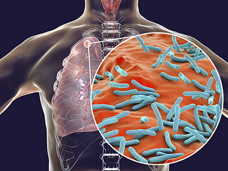
 This transport system may be widespread across many Gram-positive bacteria that contain proteins in the WXG100 superfamily. Tuberculosis kills 1 million people each year.Six years ago, Michael Niederweis, Ph.D., described the first toxin ever found for the deadly pathogen Mycobacterium tuberculosis. This toxin, tuberculosis necrotizing toxin, or TNT, became the founding member of a novel class of previously unrecognized toxins present in more than 600 bacterial and fungal species, as determined by protein sequence similarity. The toxin is released as M. tuberculosis bacteria survive and grow inside their human macrophage host, killing the macrophage and allowing the escape and spread of the bacteria.
For 132 years, the lack of an identified toxin in M. tuberculosis had contrasted with nearly all other pathogenic bacteria whose toxins contribute to illness or death. M. tuberculosis infects 9 million people a year and kills more than 1 million.
Now, in another groundbreaking work, the University of Alabama at Birmingham researcher and colleagues describe how two small ESX proteins made by the M. tuberculosis bacteria mediate secretion of TNT by pore formation in the membranes that envelop the bacteria. This finding may have broad application because a distinctive three-amino acid motif found on EsxE and EsxF — tryptophan/any-amino-acid/glycine, known in shorthand as WXG — is also found on many other small mycobacterium proteins and on the large WXG100 superfamily of bacterial proteins that resemble EsxE and EsxF.
“Here, we show for the first time that small Esx proteins of the WXG100 family have an important molecular function inside the Mtb cell by mediating toxin secretion,” said Niederweis, a professor in the UAB Department of Microbiology. “Our results suggest a dynamic mechanism of pore formation by small Esx proteins that might be applicable to other members of the large WXG100 protein family. Thus, our study not only represents a major advancement in our understanding of secretion of TNT and likely of other proteins in M. tuberculosis, but also describes a biological function for Esx-paralogs in M. tuberculosis and their homologs in the large WXG100 protein family in Gram-positive bacteria.”
TNT is one of two domains in the M. tuberculosis outer membrane protein CpnT; activity of the TNT domain of CpnT in the cytosol of the macrophage induces macrophage death by hydrolyzing NAD+. M. tuberculosis has an inner membrane and an outer membrane, and a protein needs to get through each layer to be secreted outside of the bacterium. How CpnT gets to the outer membrane was unknown.
EsxE and EsxF are part of the same gene segment as CpnT, and the UAB researchers hypothesized that the two small proteins might be involved in secretion of the toxin.
By creating different strains that lacked either EsxE or EsxF, they showed that both proteins were necessary for the translocation of CpnT to the cell surface of M. tuberculosis and for the secretion of TNT into the cytosol of macrophages infected with M. tuberculosis. Furthermore, EsxE and EsxF are surface-accessible proteins on M. tuberculosis as a membrane-associated complex.
To learn more about the mechanism of that translocation, the UAB team made mutants of each Esx protein, where the tryptophan amino acid of the single WXG motif on each protein was replaced by the amino acid alanine. The mutants showed that an intact WXG motif on EsxE and on EsxF were required for efficient CpnT translocation to the outer membrane of M. tuberculosis and subsequent TNT secretion into the cytosol of infected macrophages.
 Michael Niederweis, Ph.D. (Photography: Steve Wood)Purification of the water-soluble EsxE and EsxF proteins showed they formed EsxE-EsxF dimers, and five of these dimers assembled into star-shaped structures, as viewed by electron microscopy. Each was about 10 nanometers across, with a 3-nanometer central pore.
Experiments with planar lipid bilayers were key to understanding the molecular function of EsxE-EsxF, as they showed that the EsxE-EsxF pores formed channels through lipid membranes.
Finally, the researchers showed that the WXG motifs were required for pore formation and membrane disruption by the EsxE-EsxF complex, and the motifs mediated assembly of functional EsxE-EsxF oligomers. This now defines a biochemical role for the previously enigmatic WXG motif.
“EsxE and EsxF constitute the first known outer membrane components mediating protein secretion in M. tuberculosis,” Niederweis said. “However, it is unlikely that EsxE and EsxF are sufficient for TNT secretion, since an energy source is required in all known bacterial protein secretion systems. Therefore, it is possible that EsxE-EsxF associate with other proteins or protein complexes to achieve CpnT export and TNT secretion.”
The UAB researchers propose two models for the transport of CpnT by EsxE and EsxF. In the first, the EsxE-EsxF heterodimers form a pore in the inner membrane, and then form another pore in the outer membrane to create transmembrane channels. “Alternatively,” Niederweis said, “the inner membrane channel is extended to span the periplasm via filament formation, and connects to EsxE-EsxF pores in the outer membrane, exposing EsxF on the cell surface. In this model, the putative EsxE-EsxF channel tunnel enables export of the CpnT polypeptide to the outer membrane of M. tuberculosis, and subsequent secretion of TNT and EsxE-EsxF.”
Co-authors with Niederweis in the study, “Pore-forming Esx proteins mediate toxin secretion by Mycobacterium tuberculosis,” published in Nature Communications, are Uday Tak and Terje Dokland, UAB Department of Microbiology.
“This work was a remarkable achievement of an outstanding graduate student, Uday Tak, who did almost all of these experiments by himself,” Niederweis said. Uday Tak obtained his Ph.D. in November 2020 and is now a postdoctoral fellow at the University of Colorado-Boulder.
This paper was selected by the editor-in-chief as a highlight and is featured on a special website called “Microbiology and infectious diseases.”
Support came from National Institutes of Health grant AI121354. Electron microscopy data analysis was performed on the UAB CHEAHA supercomputer platform, which is supported by National Science Foundation grant OAC-1541310.
At UAB, Niederweis holds the Endowed Professorship in Bacteriology.