
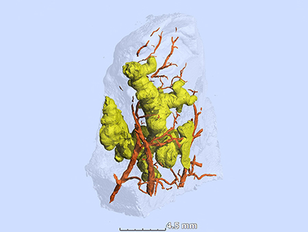
 Three-dimensional segmentation of ginger-root shaped necrotic lesions (yellow) and surrounding vasculature (red) within a sample of Mycobacterium tuberculosis-infected human lung tissue (transparent surface).
(Photography: Africa Health Research Institute)For 70 years, clinicians thought they knew the shape of tuberculosis granulomas in the lungs of patients. Histology — the study of microscopic structures in thin slices of lung tissue in the 1940s and 1950s — showed round features, and researchers intuitively assumed that meant the granulomas were spherical or ovoid.
That long-lived paradigm is now shown to be wrong, in a study by researchers at the University of Alabama at Birmingham and the Africa Health Research Institute, or AHRI, Durban, KwaZulu-Natal, South Africa. The historical histology, essentially, was a two-dimensional look at structures, similar to cutting a very thin slice through a tree branch, a slice that would look round or oval.
The new research has created a three-dimensional view of diseased lung tissue from tuberculosis patients, using micro-computed tomography, or microCT. This revealed that the larger granulomas were anything but round — rather they had complex, branched shapes. One granuloma looked somewhat like a ginger root, another like a cluster of early buds on a cherry tree, before the blossoms appear. The tuberculosis granulomas showed marked heterogeneity in shape, volume and number in the lung sections. (See this study link for videos of the three-dimensional views.)
A granuloma is an aggregation of immune cells summoned in response to chronic inflammation. In tuberculosis, the granulomas are often necrotic, meaning they are a mass of formless dead debris.
Researchers led by Adrie Steyn, Ph.D., a UAB professor of microbiology and a member of the AHRI, used microCT, histology and immunohistochemistry to construct three-dimensional views of necrotic granulomas, and also vasculature and airways. This yielded an unanticipated insight into the spatial organization of tuberculosis granulomas in relation to airways and vasculature.
“Unlike depictions of granulomas as simple spherical structures,” Steyn said, “human necrotic granulomas exhibit complex, cylindrical, branched morphologies that are connected to the airways and shaped by the bronchi.”
Steyn says these visualizations have three impacts. They highlight the likelihood that a single structurally complex lesion could be mistakenly viewed as multiple independent lesions when evaluated in two dimensions. Second, the lack of vascularization within obstructed bronchi establishes a paradigm for anti-mycobacterial drug tolerance, since the microbes are protected from a full delivery of blood-borne bactericidal drugs. Third, the results suggest that a bronchogenic spread of the Mycobacterium tuberculosis pathogen re-seeds the lung.
“Hence, our findings provide a rationale for considering aerosolized anti-tuberculosis drug delivery,” Steyn said. “This approach could allow delivery of high, local concentrations of drugs directly into granulomas or cavities in the lung, possibly reducing treatment times, systemic dosing and toxicity.” Infections by the Mycobacterium tuberculosis bacterium kill about 1.3 million people a year.
Steyn and colleagues say, to their knowledge, this is the first three-dimensional description of granulomas, airways and vasculature for any bacterial pulmonary pathogen.
 Adrie Steyn, Ph.D., a UAB professor of microbiology and a member of the AHRI.
(Photography: Steve Wood)Infected human lung tissue is readily available for study in Durban because that part of Africa is an epicenter for tuberculosis. The Inkosi Albert Luthuli Central Hospital, not far from AHRI, does resection surgeries to remove diseased lobes from tuberculosis patients. MicroCT is similar to the common medical CAT scans but has a much finer resolution. The researchers used resolutions as small as 12 microns, which is about the thickness of a sheet of plastic kitchen wrap. Research leader Steyn maintains labs at UAB and the AHRI. He lives in Durban and visits UAB about six times a year.
The study, “µCT Analysis of the Human Tuberculous Lung Reveals Remarkable Heterogeneity in 3D Granuloma Morphology,” was published in the American Journal of Respiratory and Critical Care Medicine. The journal also published an editorial on the Steyn study.
Co-authors with Steyn are Gordon Wells, Kievershen Nargan and Kapongo Lumamba, AHRI; Joel N. Glasgow, UAB Department of Microbiology; Rajhmun Madansein and Kameel Maharaj, Inkosi Albert Luthuli Central Hospital and University of KwaZulu-Natal, Durban, South Africa; Robert L. Hunter, University of Texas Health Sciences Center at Houston; Threnesan Naidoo, Inkosi Albert Luthuli Central Hospital; and Llelani Coetzer, Stephan Le Roux and Anton du Plessis, Stellenbosch University, South Africa.
Support came from National Institutes of Health grants Al111940, AI134810, AI137043, AI138280 and A127182; a Bill and Melinda Gates Foundation Award; and pilot funds from the UAB Center for AIDS Research, the UAB Center for Free Radical Biology, and the Infectious Diseases and Global Health and Vaccines Initiative. Support also came CRDF Global, the South African Medical Research Council and a South Africa National Research Foundation BRICS Multilateral grant.