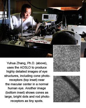
New Views Inside the Eye
By Tyler Greer
 Fifteen years ago, Yuhua Zhang, Ph.D., was learning to design cameras, telescopes, and microscopes in his native China. Then his mother-in-law developed sudden, severe bleeding in her left eye, and his focus changed. After learning that doctors did not have the equipment to produce high-resolution images of the retina, he devoted his career to ocular imaging. Now, Zhang, a UAB assistant professor of ophthalmology, has developed a high-resolution imaging instrument that provides an unequaled view of the human eye.
“This is, to our knowledge, the fastest practical adaptive optics for the living human eye,” Zhang says. “The development of this instrument has positioned UAB at the forefront of this emerging technology—available at only five other centers worldwide.”
Adaptive optics is technology that was originally created to help high-powered telescopes see clearly through the turbulent atmosphere in deep space. Applied to vision, adaptive optics enables retinal imaging systems to compensate for the optical defects of the human eye’s cornea and lens, offering the ability to visualize living cells within the eye.
UAB’s adaptive-optics scanning-laser ophthalmoscope (AOSLO) will help ophthalmologists detect age-related macular degeneration, diabetic retinopathy, and glaucoma, allowing them to treat the diseases earlier and slow their progression.
UAB’s AOSLO is the third generation of the device. Zhang completed an earlier version while working at the University of California at Berkeley with optometry professor Austin Roorda, Ph.D., but its clinical use was limited. When Zhang set up his UAB lab in 2008, his goal was to develop a new AOSLO capable of high dynamic range, ocular-aberration compensation, and high-speed imaging acquisition.
“The images are of unprecedented quality, enabling video images of the microscopic blood flow and photoreceptors for diagnosis of eye diseases,” Zhang says. The device can show well-resolved cone photoreceptors in the macula, the retina’s central region housing the densest pack of cones, the cells forming the fine and color vision. “In particular, it can image the rod photoreceptors in the living, awake human eye, a breakthrough that will improve our understanding of rod-mediated blinding diseases such as age- related macular degeneration, retinitis pigmentosa, and cone-rod dystrophy,” Zhang says. “We are one of only three labs in the world that currently possess this ability.”
The AOSLO can even project clear visual stimuli onto the retina to facilitate testing of retinal function. “We will be able to diagnose diseases at an earlier stage and provide sensitive evaluation of the treatment efficacy at the cellular level,” Zhang says.
Initially, Zhang will use the AOSLO to study age-related macular degeneration and primary open-angle glaucoma with UAB ophthalmologists Cynthia Owsley, Ph.D.; Christopher Girkin, M.D.; Christine Curcio, Ph.D.; and Douglas Witherspoon, M.D. “We also will study other common medical and neurologic conditions, including hypertension, diabetes, multiple sclerosis, and Alzheimer’s disease,” Zhang says.
—This article originally appeared in the summer 2011 issue of UAB Medicine magazine.