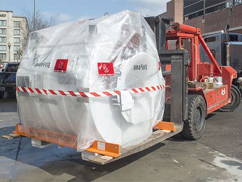
 The PET/MR, being delivered to UAB, is a tremendous advancement in technology that will fit exceptionally well with UAB's PET/CT machines and cyclotron. The next closest place in the U.S. using this technology for standard-of-care imaging is in North Carolina.Sixteen months after the installation of a groundbreaking GE Signa PET/MR imaging machine to accelerate numerous research efforts at the University of Alabama at Birmingham, clinicians will now be afforded the opportunity to use it in a whole new way — for standard-of-care clinical PET imaging.
The ability to now use the PET/MR machine for clinical work puts UAB in rarified air; although there are approximately 50 research PET/MRs in the United States, there are very few programs using them for clinical work. The closest place in the United States using PET/MR technology for standard-of-care imaging is in North Carolina.
“The ability to use this extraordinary machine for clinical care truly sets UAB apart from other academic medical institutions in the Southeast, the United States and, really, worldwide,” said Cheri Canon, M.D., the Witten-Stanley Endowed Chair of Radiology in UAB’s School of Medicine. “There are not many with this capability. And the addition of PET/MR to go along with the other machines available in our Advanced Imaging Facility enables us to provide our patients an additional and a truly unique, cutting-edge tool — one that has great potential as we move toward precision medicine.”
PET/MRI combines two complementary advanced imaging techniques — positron emission tomography (PET) and magnetic resonance imaging (MRI) — into a single scanner that can acquire PET and MRI data simultaneously. The simultaneous acquisition of PET and MR data enables new opportunities for physicians who need diagnostic imaging of patients with cancer, brain disorders and heart disease, among other diseases and neurological issues.
PET uses small amounts of radioactive drugs to measure a wide range of important biological processes, including blood flow, metabolism and biochemistry, while MRI provides high-resolution structural images as well as important functional information such as heart motion and brain activity.
 Jonathan McConathy, M.D., Ph.D., director of UAB’s Division of Molecular Imaging and Therapeutics.“When you combine these two innovative tools, clinicians may be able to see early cellular changes before any anatomical changes could be observed and can then fuse the anatomical images and active biochemistry data together to pinpoint the area of abnormal cell growth,” said Jonathan McConathy, M.D., Ph.D., director of UAB’s Division of Molecular Imaging and Therapeutics. “Both of these imaging modalities are very good at what they do, and combining them gives physicians very sophisticated, biologically specific imaging along with high-resolution characteristics. It’s a tremendous advancement in technology that will fit exceptionally well with our PET/CT machines and cyclotron.”
PET/MRI also offers several important advantages over other imaging modalities. For example, PET/MRI can reduce radiation exposure by using MRI instead of computed tomography, which is currently used in the vast majority of clinical PET studies with PET/CT. Radiation reduction is particularly important for children and young adults who will undergo multiple PET studies during the course of their treatment
Patients who need both PET and MR imaging also have the ability to have both done within the same imaging session rather than requiring two separate appointments.
Radiologists and nuclear medicine physicians interpreting the study have the PET and the MRI studies to review at the same time, which is more efficient and has the potential to improve diagnostic accuracy.
Finally, MRI is particularly well-suited for certain types of studies, including imaging of the brain, heart, liver, pelvis and bone marrow. For some patients undergoing imaging of these regions of the body, PET/MRI may be preferred over PET/CT.
Advanced Imaging Facility advantages
UAB’s Advanced Imaging Facility is the Southeast’s most advanced and can engage a range of disciplines for research and patient-care endeavors — from scientists and physicians in UAB’s Comprehensive Cancer Center, the neurosciences and cardiology, to high-level mathematicians, bioengineers, vision scientists and others.
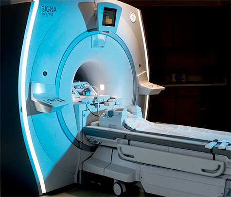 UAB’s SIGNA PET/MR, an innovative machine from GE, combines two complementary advanced imaging techniques — positron emission tomography (PET) and magnetic resonance imaging (MRI) — into a single scanner that can acquire PET and MRI data simultaneously and help physicians pinpoint areas of abnormal cell growth.In addition to the PET/MR, the facility houses two PET/CT machines and a cyclotron, a particle accelerator that moves protons along a spiral path to strike a target. UAB uses its cyclotron to make medical imaging agents for clinical and research applications. It is also capable of making agents for therapy.
One way it is believed PET/MR will play an important role in standard clinical care is through the delivery of these specially made medical imaging agents produced by the cyclotron.
Many PET studies use short-lived radioisotopes, or medical imaging agents, that can last for 20 to 110 minutes. Some of these agents are made routinely by radio pharmacies, but it is helpful for facilities engaged in high-level research and clinical protocols to make the agents on-site.
“That’s where the cyclotron comes into play in all of this because using it with PET/MR gives us options to make new or known research tracers here that aren’t available except at a few sites,” McConathy said. “Then, we can use these tracers with PET/MR or PET/CT.”
While patients often get PET and MRI separately and scientists fuse the images to simulate the PET/MR capabilities, acquiring all of the data from the imaging at one time, through one scan, will help physicians be more precise in targeting affected areas.
“When we are able to acquire all of the data in one scan, I think everyone involved does a better job diagnostically with the patient,” McConathy said. “And in many ways, the medical community is still learning about PET/MR, what’s it capable of and how it fits with PET/CT. PET/MR is a new and important tool, but it’s not one that’s going to replace PET/CT. But for some patients, especially ones where we can speed up their diagnosis, have them come for one imaging study instead of two, or in instances where we can reduce radiation, those are obvious areas to use PET/MR right now. We’re excited to see where these opportunities will take us in the future in patient care and research.”