By Hannah Buckelew
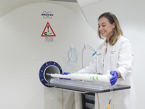
The Department of Biomedical Engineering boasts a diverse research portfolio covering a wide range of topics. Biomedical imaging, for example, is an essential tool in visualizing the body’s internal organs. To showcase the impactful contributions of imaging research in the department, Anna Sorace, Ph.D., an associate professor in the Departments of Biomedical Engineering and Radiology, and director of UAB’s Small Animal Imaging Facility, answers a series of questions.
Sorace has expertise in multimodality, non-invasive quantitative imaging to improve detection, monitoring and therapy of cancer in both preclinical animal models and clinical cancer research. The Sorace Research Lab is focused on improving cancer patient care through translational advancements in cancer imaging.
In your own words, what is biomedical imaging?
Biomedical imaging is an approach that uses medical imaging instrumentation to provide a noninvasive approach to understand anatomical, functional, and molecular processes in normal physiology and diseases.
How did you get involved imaging?
My initial research interest started in cancer drug delivery, and I quickly realized that imaging provides the quantitative data necessary to help guide therapeutic interventions to get temporal and spatial information.
How does imaging intersect in BME and radiology?
BME has always had imaging as a central component of education and research. The field of radiology is continuously advancing both in instrumentation and image analysis approaches and is moving from being primarily diagnostic imaging to include theragnostic and prognostic imaging approaches in medicine. These advancements provide the opportunity to intersect more computational strategies into imaging research, which pairs nicely with BME.
Can you describe some of your research specifically with imaging in breast cancer?
Our goal is to better personalize cancer treatment by using computational imaging strategies to understand the tumor microenvironment and the variations, or heterogeneity, within a tumor. Because every tumor is different, imaging provides a 3D approach to quantify key components such as the vasculature and immune cells to help identify how that cancer will respond to various combinations of therapy. This allows the development of predictive biomarkers of response and approaches for image-guided therapeutic interventions.
How is imaging impacting the field of BME? UAB Hospital as a whole?
Imaging intersects and is used by every department within UAB Hospital! As imaging advancements are needed clinically, it will continue to grow as an integral component of BME.
What are your hopes for the future of imaging at UAB?
My goal for imaging at UAB is to make quantitative imaging approaches more accessible to every researcher, therefore creating strategies that allow non-imaging scientists or engineers to incorporate imaging into their research and answer their unique scientific hypotheses.