by Ross Hansen, MD
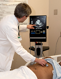
Point-of-care ultrasound or (POCUS) has emerged over the years as a valuable tool for clinicians to quickly triage patients at the bedside and reduce the time to medical intervention. The use of ultrasonography has long been used by colleagues in radiology and emergency medicine, particularly in the use of standard-of-care focused assessment with sonography for trauma (FAST) examinations. However, only recently has ultrasound become more commonplace in other clinical contexts such as the medical wards including medical and cardiac critical care medicine.
Notably, point-of-care ultrasound has become a useful tool in improving the precision of bedside procedures, triaging undifferentiated shock, and assessing a patient’s fluid status. When used correctly, POCUS can assist providers with arriving at the correct diagnosis in a shorter period of time or deciding medical management quickly. A 2004 study found a nearly 30% increase in number of physicians who were able to correctly identify non-traumatic, symptomatic hypotension in a 15 minute period via the use of skilled but simple ultrasound techniques. The idea being that a simple view of the heart with an ultrasound probe can help quickly triage patients with shock. Despite all of its conveniences, the results from POCUS can vary widely among providers and requires a foundation of medical and technical understanding. Providers may find that they have adequate medical background but not necessarily the technical skill to guide management of patients.
Bridging this technical “gap” is a relatively new topic of discussion within internal medicine residency curriculums and will become a valuable skill set for current trainees as they begin to think about entering practice. We're excited UAB has begun to introduce new ultrasound training opportunities for residents beginning with the 2018-2019 intern class that will involve dedicated time with critical care faculty practicing diagnostic ultrasound. The curriculum is headed by faculty members within the Department of Medicine, Critical Care and Cardiology, Drs. Robert Smola, Micah Whitson and Sam McElwee.
An afternoon seminar allows residents to work in a small group with a critical care attending in the UAB Hospital simulation center. Residents are able to review the basic technical aspects of ultrasound, including identifying anatomic windows, adjusting the image with the orientation of the probe, as well as changes to the image depth and gain on the monitor. Residents then take to simulation where they can work together to practice their skills troubleshooting an image and developing a differential diagnosis for several cardiac and pulmonary cases. In the presence of an attending, residents are able to get direct feedback as they work through each of these clinical scenarios. In doing so, residents can begin to translate their learned ultrasound skills in the simulation lab to the bedside.
When speaking with Dr. Smola, he explained the larger goals of the curriculum are to improve quality and safety outcomes of our procedures as well as appropriately influence our decisions on the wards. Residents have also had the opportunity over the past several years to rotate with the hospitalist procedure service which has allowed for earlier exposure to procedural point-of-care ultrasound allowing residents to practice a variety of ultrasound guided procedures. Getting exposure to both of these experiences is an excellent opportunity for UAB residents to become increasingly comfortable with ultrasound prior to implementing their skills on the ward and eventually in practice.
Reference: Jones, A., Sullivan, M. & Kline, J. (2004). Randomized, controlled trial of immediate versus delayed goal-directed ultrasound to identify the cause of nontraumatic hypotension in the emergency department patients. Critical Care Medicine, 32(8)