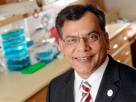
 Keshav K. Singh, Ph.D., is a professor of genetics in the UAB School of Medicine. Patients with colon and rectal cancer have somatic insertions of mitochondrial DNA into the nuclear genomes of the cancer cells, University of Alabama at Birmingham researchers report in the journal Genome Medicine.
This study provides the novel role for mitochondria inducing genomic instability in the nucleus of cancer cells, the UAB researchers say.
Overall, a study of the genome sequences of 57 colorectal cancers showed, on average, 4.42-fold more somatic nuclear mitochondrial DNA as compared to matched healthy blood controls. This suggests that migration of mitochondrial DNA into the nuclear genome plays an important role in development of cancer, say corresponding authors Hemant K. Tiwari, Ph.D., and Keshav K. Singh, Ph.D. Tiwari is professor of biostatistics in the UAB School of Public Health, and Singh is professor of genetics in the UAB School of Medicine.
Numerous mitochondrial organelles reside in nearly all higher eukaryotic cells, and they function as the powerhouses of the cells to create ATP, the cell’s major energy source. Human mitochondria have just 37 genes. The mitochondria are located in the cytosol of the cells, outside of the cell nucleus; the cell’s DNA genome is located inside the nuclear membrane.
Over eons, pieces of mitochondrial DNA have naturally inserted into eukaryotic genomes; at birth, for example, humans have between 755 and 1,155 germline mitochondrial DNA inserts that have been passed on through generations. These germline, inherited mitochondrial DNA insertions are seen over a wide range of organisms, including humans, plants, yeast, malaria parasites and nematodes.
Besides DNA, entire mitochondria, mitochondrial proteins and mitochondrial RNA can also be found in the nucleus; but their roles in human health and disease remain relatively unexplored. The increased mitochondrial DNA in the genomes of the cancer cells observed by the UAB team suggests somatic insertion of mitochondrial DNA occurred as the cancer developed.
The median nuclear mitochondrial DNA fold-change in colorectal tumors of women was higher than the median for men. There appeared to be a correlation of increased fold-change with poorer patient survival, but the researchers say further analysis of a larger sample size is needed to make a definite conclusion.
| Besides DNA, entire mitochondria, mitochondrial proteins and mitochondrial RNA can also be found in the nucleus; but their roles in human health and disease remain relatively unexplored. The increased mitochondrial DNA in the genomes of the cancer cells observed by the UAB team suggests somatic insertion of mitochondrial DNA occurred as the cancer developed. |
The somatic mitochondrial DNA inserts appeared to correlate with gene-rich segments of the genome, and the researchers mapped hotspots on the mitochondrial genome for migration into the nuclear genome.
Since the gene product of YME1 is a potent suppressor of mitochondrial DNA migration into the genome of the yeast Saccharomyces cerevisiae, Tiwari and Singh investigated the human homologue of the YME1 gene, called YME1L1. Both gene products have conserved roles in mitochondrial assembly (mitochondria are made up of 2,000 proteins encoded by the nuclear genome and 13 encoded by the mitochondrial genome), mitochondrial integrity and DNA metabolism. However, a role for the human YME1L1 in suppressing migration of mitochondrial DNA into the nucleus was unknown.
As indirect evidence, the UAB researchers found that 16 percent of the 57 colorectal cancer sequences examined had mutations in the human gene YME1L1. An expanded analysis of colorectal cancer sequences in The Cancer Genome Atlas database showed a high incidence of YME1L1 mutations. The researchers also found high mutation rates of YME1L1 in other cancers.
The UAB team next showed that knocking out YME1L1 in a human cell line strikingly increased the amount of mitochondrial DNA in the nuclear fraction of YME1L1 knockout cells. These results identified YME1L1 as the first reported suppressor gene for nuclear migration of mitochondrial DNA in humans, and they suggested that inactivation of YME1L1 leads to increased migration of mitochondrial DNA into the nucleus.
This was tested in yeast. A marker that can only be transcribed in the nucleus was inserted into yeast mitochondrial DNA. When the yeast YME1 was mutated, the marker was 20-fold elevated compared to yeast with non-mutated YME1. When human YME1L1 was introduced into yeast with the mutated YME1, the human gene product partially rescued the YME1 mutation, preventing migration of mitochondrial DNA into the nucleus. Thus, human YME1L1 appears to suppress migration of mitochondrial DNA into the nucleus.
Tiwari and Singh have dubbed a new term — numtogenesis (pronounced “noo-mitto-genesis”) — for this phenomenon of mitochondrial occurrence in the nucleus.
In a companion paper in Analytical Biochemistry, the Singh laboratory and the Bernd Friebe laboratory at the Department of Plant Pathology, Kansas State University, describe a molecular tool to rapidly detect and analyze insertion of mitochondrial DNA into the genomes of cells.
Up to now, analysis of mitochondrial DNA inserted into the nuclear genome depended on deep genome sequencing combined with comprehensive computational analysis of the entire genome, a time-consuming, cumbersome and cost-prohibitive approach. The researchers, led by corresponding author Singh and first author Dal-Hoe Koo, research assistant professor at Kansas State, developed a single-molecule mitochondrial Fiber-FISH technique, using fluorescence in situ hybridization. This permits high-resolution mapping of genes and chromosomal regions on single fibers of DNA, and it targets the physical location of mitochondrial DNA probes down to a resolution of 1,000 base pairs.
“This novel technique,” they said, “should help distinguish and monitor cancer stages and progression, aid in elucidation of basic mechanisms underlying tumorigenesis, and facilitate analyses of processes related to early detection of cancer, screening and/or cancer risk assessment.”
Besides Tiwari and Singh, co-authors of the Genome Medicine paper, “Migration of mitochondrial DNA in the nuclear genome of colorectal adenocarcinoma,” are Vinodh Srinivasainagendra, Michael W. Sandel, Bhupendra Singh, Aishwarya Sundaresan, Ved P. Mooga and Prachi Bajpai, UAB departments of Biostatistics and Genetics.
Funding came from Veterans Affairs grant 1I01BX001716, a UAB NCTN–LAPS Program Translational Research Award, and National Institutes of Health grant HL072757.
Besides Singh, Friebe and Koo, co-authors of the Analytical Biochemistry paper, “Single-molecule mtDNA Fiber FISH for analyzing numtogenesis,” are Jiming Jiang, University of Wisconsin-Madison; Bikarm S. Gill, Kansas State University; Paul D. Chastain, William Carey University, Hattiesburg, Mississippi; and Bhupendra Singh, Upender Manne and Hemant K. Tiwari, UAB.
Funding came from National Institutes of Health grant CA204430, Veterans Affairs grant 5I0I BX001716, UAB Cancer Center LAPS-NCTN pilot project grant, and a National Science Foundation contract 1338897 to the Wheat Genetics Resources Center, Kansas State University.
At UAB, Tiwari holds the William “Student” Sealy Gosset Endowed Professorship in Biostatistics, UAB School of Public Health, and Singh holds Joy and Bill Harbert Endowed Chair in Cancer Genetics.