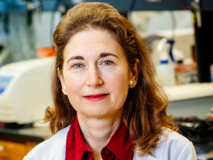
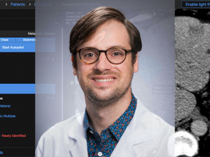
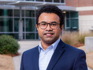
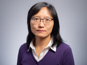
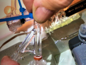
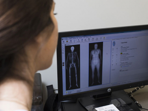
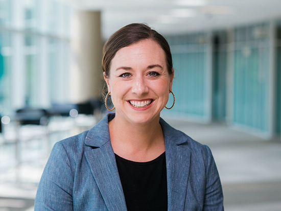
 Anna Sorace, Ph.D., director of UAB's Small Animal Imaging Facility and associate professor in the Department of Radiology, has leveraged HSF-GEF grants to help grow UAB’s nuclear imaging program.At one time, immunology was only for immunologists. Now “the world of immunology has infiltrated almost every disease,” said Anna Sorace, Ph.D., associate professor in the Department of Radiology and director of the UAB Small Animal Imaging Facility. “Everyone has to have a minor in immunology these days, and everyone needs an immunologist to collaborate with for their research,” said Sorace, who also has an appointment in the Department of Biomedical Engineering.
UAB’s growing nuclear imaging program paired with the Cyclotron Facility is a national leader in the development of “immunoPET and related immuno-imaging,” Sorace said. The major advantage of these modalities is that they allow for longitudinal imaging of conditions over time, she notes. UAB researchers create radioactive elements in the university’s cyclotron and then pair them with biological markers that can bind to macrophages, T cells and other key players in the immune system. The radioactive elements create signal for PET images.
But before these promising imaging probes can reach patients, they must meet a host of regulatory and experimental requirements. “You have to prove that the target you have found is what you are targeting and it is in fact there,” Sorace said. “If you are looking specifically at an M2 macrophage, you need to show that there are similar populations” in the tissue taken from an animal that matches the imaging of that animal.
The trouble is that the tissue samples are radioactive after imaging is done. “The imaging facility did not have the ability to take those samples to UAB’s well-equipped Flow Cytometry Core,” Sorace said. “We wanted to biologically validate this non-invasive imaging, which is critical to advance these novel tests.”
“We wanted to biologically validate this non-invasive imaging, which is critical to advance these novel tests.”
The NIH and NSF have programs to fund new or cutting-edge equipment, but “buying a flow cytometer for an imaging core would not be a fundable NIH S10,” Sorace said, referring to the NIH equipment grant mechanism. Especially because the one she wanted was the exact same model already found in the Flow Cytometry Core in order to easily pair together ongoing research. Instead, Sorace successfully applied for funding through UAB’s unique Health Services Foundation-General Endowment Fund grants program in 2021. That funding, paired with additional support from the departments of radiology and chemistry and the O’Neal Comprehensive Cancer Center, allowed Sorace’s facility to add the new equipment.
“It really allows us to expand our infrastructure and utilize the expertise in the Flow Cytometry Core,” Sorace said. “This equipment is matched to one of their instruments, so if someone wants to be trained, they can implement that in the Flow Cytometry Core and then use those protocols for pairing with nuclear imaging in the Small Animal Imaging Facility.”
“These grants bring a lot of groups together to talk about the gaps that we need to fill to be stronger as an institution. They have greatly enhanced the infrastructure in our core facility.”
Now, with the flow cytometer in place, “we can do these studies from start to finish, with matched imaging and biological assays,” Sorace said.
Sorace’s core supports the O’Neal Comprehensive Cancer Center, the Heersink School of Medicine, the O’Brien Center for Acute Kidney Research and neuroscience researchers, as well as chemistry and engineering. Investigators can do imaging through the core that is assisted or unassisted, Sorace says. “From a core facility perspective, I want to create an environment that will allow people to implement imaging and imaging research into their own disease studies, without having an imaging background,” she said. “It is a full-service approach; we can take things from start to finish. We can also do image analysis and then autoradiography, which is similar to immunohistochemistry for radioactive contrast agents. Flow cytometry is that next piece of the puzzle.”
In late 2023, Sorace learned that she had been funded for a second HSF-GEF grant, which will support expansion of preclinical PET imaging in the Small Animal Imaging Facility. “These grants bring a lot of groups together to talk about the gaps that we need to fill to be stronger as an institution,” Sorace said. “They have greatly enhanced the infrastructure in our core facility.”
How do the HSF-GEF awards advance UAB? Faculty explain their projects:
How the HSF’s General Endowment Fund awards help UAB compete on a national stage
Advancing faculty careers through the UAB Academy of Health Professions Educators
Building immuno-imaging at UAB through specialized technology
Turning a pilot project into the standard of care
Making the leap from one hospital to another through UAB’s STEP Program
Building a health system that can learn calls for “team science to the max”
Using geographic information systems in patient-oriented research
