The Department of Pathology at UAB makes thousands of diagnoses for patients each year. Of an estimated 150,000 annual visits to UAB Hospital, 90% of patients require some form of pathology service. Timely and accurate diagnoses are critical to determine the best treatment options, and yet reaching those diagnoses can be a very delicate and complex process. Cristina Magi-Galluzzi, M.D., Ph.D., Robert and Ruth Anderson Endowed Chair in Pathology, alongside skilled clinicians in the Department of Pathology, showcases the journey of a pathology specimen to reach diagnosis.

It all starts here
The specimen begins its journey toward a diagnosis once delivered to our accessioning team. It is established in our database, appropriately labeled, and matched with relevant patient history before being taken to the macroscopy laboratory.
In the macroscopy laboratory, our skilled team of Pathologists Assistants and trainees work to properly section and analyze the specimen, taking note of its dimensions and characteristics. Once representative sections of the specimen have been submitted into appropriate cassettes, they must be fixed in formalin and processed in various chemical solutions for 8 to 12 hours.
From biopsy to glass slide
Once the specimen has been processed in solution, it is embedded into a wax block, which allows the specimen to be thinly sliced by trained laboratory professionals to create glass slides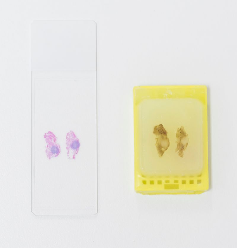 Glass slide (left) and paraffin wax block for pathologists to examine. The thin wax pieces must be stained with Hematoxylin and Eosin to make its properties visible under the microscope.
Once stained, the case turn out team organizes the glass slides into folders with its correct case information. When all the samples from the specimen are ready and the slides are properly organized, the case is assigned to a pathologists' subspecialty box. A trainee in that specialty's rotation will preview the case before assigning it to its specified faculty attending.
Standard H&E tissue stains show the overall shape and structure of cells. Frequently, faculty will need to order additional testing necessary to make the most accurate diagnostic interpretation.
The need for additional testing
Our immunohistochemistry laboratory goes one step further by performing IHC testing essential in diagnosing cancer type and biomarker expression. This test generally takes an additional 1 to 2 days to complete, depending on the volume.
Other tests that may likely be ordered are molecular testing, which require DNA and RNA extraction from the sample. Molecular testing can be used for diagnosis both in cancer or infectious disease, or to guide treatment.
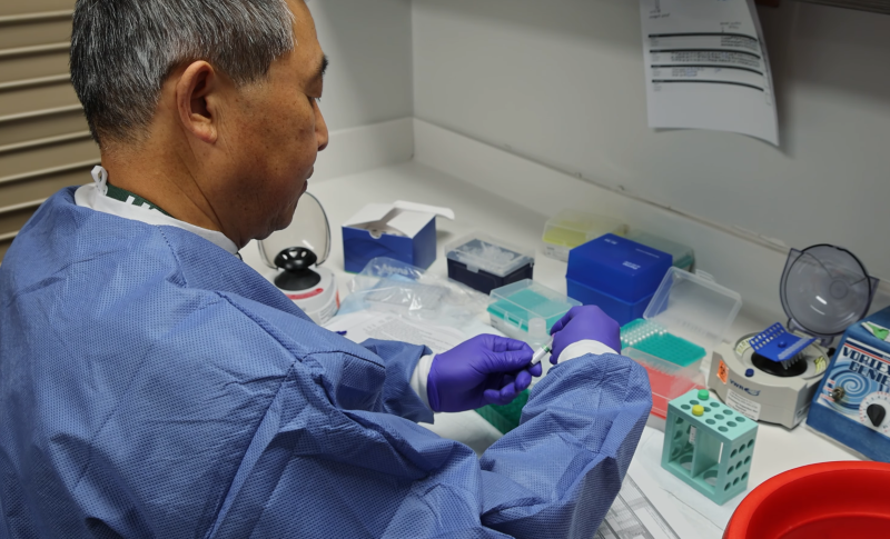 Ximeng Liu preparing samples for molecular testing
Constructing the final report
The final step in the specimen's journey is its pathology report construction and verification. Once all test results are available, the case is reviewed again by an attending with subspecialty expertise to determine and verify the diagnosis and complete the report, which clinicians will have access to and share with the patient.
"Our patients are the driving force behind our commitment to diagnostic excellence," said Magi-Galluzzi. "Behind every specimen is a person, a story, and a journey- and we never lose sight of the human being behind the diagnosis. Every case we sign out carries a responsibility that we hold deeply and never take for granted. We are proud to care for our patients in Birmingham and beyond and honored to be your pathologists."