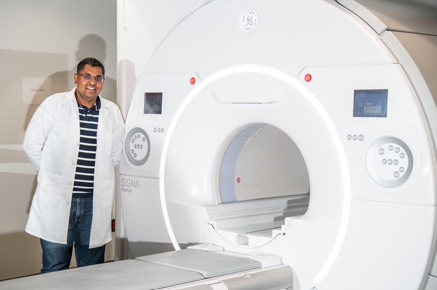
Magnetic Resonance Imaging (MRI) is a powerful research tool helping scientists answer important questions about how the body works, how diseases develop, and how new treatments can improve lives. From understanding how the brain and body function and develop to studying diseases affecting the brain, lungs, heart, liver, kidneys, and muscles, the UAB Research MRI Core helps researchers visualize the human body in remarkable detail without surgery or radiation.
MRI uses powerful magnets and radio waves to create highly detailed images of the body's organs and tissues. Unlike X-rays or CT scans, MRI does not use ionizing radiation, making it a valuable and widely used tool for both clinical care and research.
“MRI empowers researchers to visualize the human body in remarkable detail, advancing our understanding of how it functions, how diseases develop, and how new treatments can improve lives, without surgery or ionizing radiation,” said Virendra Mishra, Ph.D., associate professor and co-director of the Research MRI Core.

The UAB Research MRI Core serves as a hub for investigators across UAB and beyond, providing access to state-of-the-art imaging technology, technical expertise, and advanced data analysis. Research projects range from developing new MRI methods and improving imaging technology to supporting large clinical studies that help answer important questions about health and disease.
The UAB Research MRI Core is also available to investigators from partnering institutions and organizations. Scientists interested in using the facility can work directly with core staff to develop a customized imaging plan, identify the appropriate scanner, and receive technical support throughout their project. This collaborative approach makes advanced MRI technology accessible to both experienced imaging researchers and investigators new to MRI-based studies.
“The UAB Research MRI Core serves investigators across UAB and beyond, supporting diverse projects, from developing innovative MRI methods and technology to advancing large nation-wide clinical and research studies through collaborative access to advanced imaging, technical expertise, and customized research support,” Mishra said.
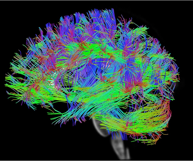
The facility features several specialized MRI systems designed for different research needs. Both flagship research scanners are equipped with advanced hardware that produces exceptionally detailed images and supports cutting-edge applications such as functional MRI, diffusion imaging, and spectroscopy. Additionally, the Core can now perform studies to observe lung integrity and also collect brain signatures, similar to functional MRI, using naturalistic conditions. Furthermore, the Core is now equipped with a mock MRI scanner to help patients and research participants acclimate to the scanning environment to reduce claustrophobia and anxiety.
“The UAB Research MRI Core features specialized, state-of-the-art MRI systems and advanced tools that support high-resolution imaging, innovative applications, naturalistic brain and lung studies, and improved participant comfort through a mock MRI scanner,” Mishra said.
Safety remains the Core's highest priority. Every participant undergoes a thorough screening process before entering the MRI environment. Because MRI uses a powerful magnetic field, staff carefully evaluate implants, medical devices, and metallic objects to ensure safe participation. Researchers and MRI technologists are specially trained to maintain rigorous safety standards and provide a comfortable experience for participants.
To help people become familiar with the MRI experience, the Core also utilizes a mock scanner that replicates the sights, sounds, and environment of a real MRI system. This educational tool helps reduce anxiety and improve participation, especially for children and first-time participants.
“Safety is the UAB Research MRI Core’s highest priority, with thorough screening, specially trained staff, and a mock scanner helping ensure a safe, comfortable, and reassuring experience for every participant,” Mishra said.
Every MRI scan contributes to a larger goal: improving our understanding of human health and disease. Whether supporting a single investigator's research question or enabling large collaborative studies, the UAB Research MRI Core helps transform ideas into discoveries, providing the technology, expertise, and innovation needed to better understand health and disease.
“At UAB, the Research MRI Core transforms innovative ideas into meaningful discoveries, empowering investigators and collaborative studies with the technology and expertise to advance our understanding of human health and disease,” Mishra said.