Pathology Core Research Lab provides state-of-the-art histology and histomorphometric analysis of various tissue types, and is specialized on bone samples. The present location on the 4th floor of Zeigler Research Building has approximately 2022 square feet of space in close proximity to many researchers who utilize our services.
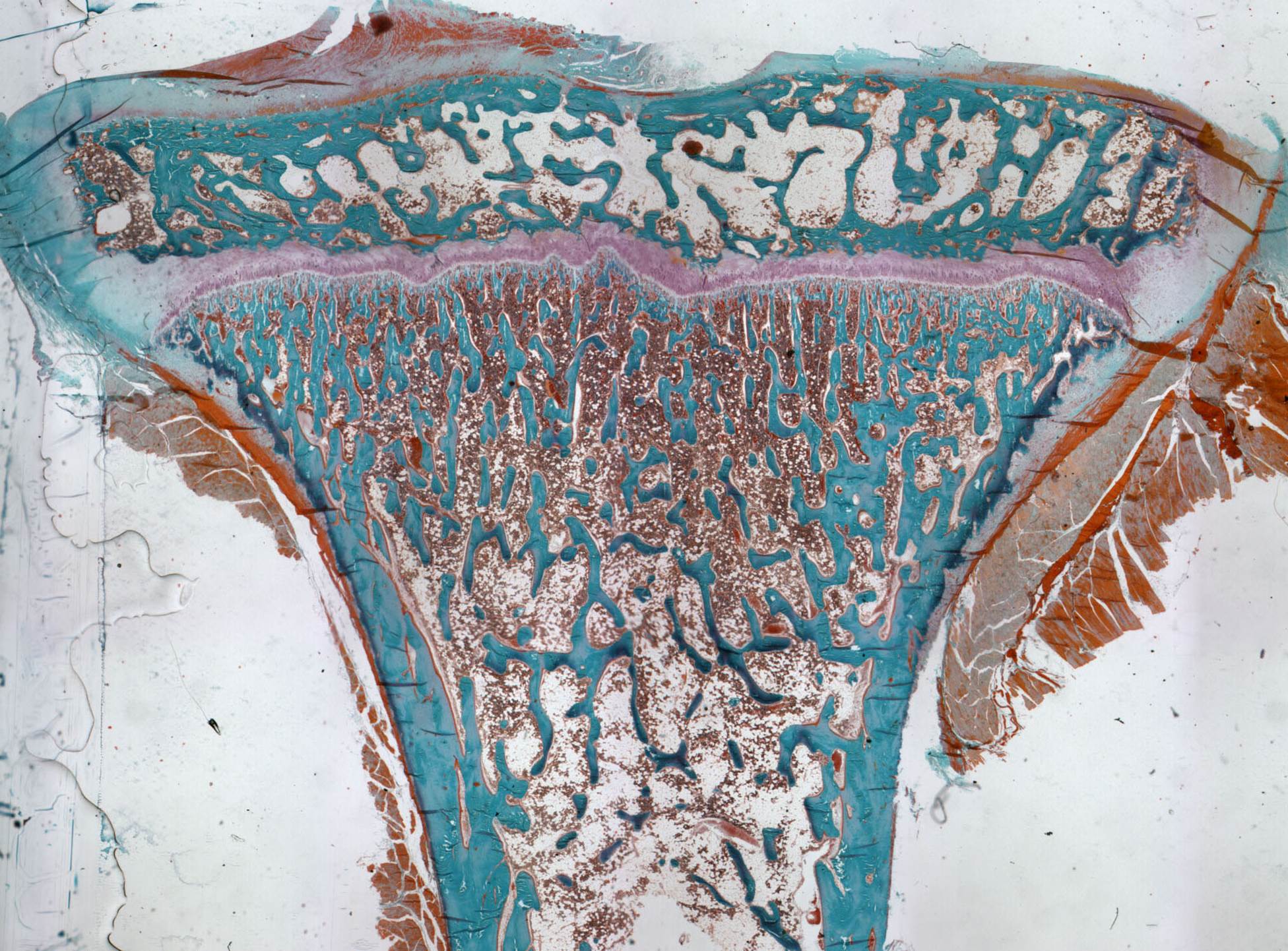
Plastic section of Monkey Tibia non-decaled section Goldner's Trichrome
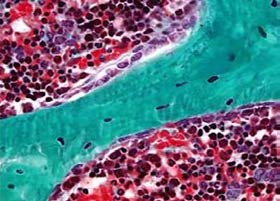
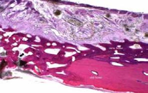
Plastic embedded bone section with Goldner's trichrome, at higher Magnification
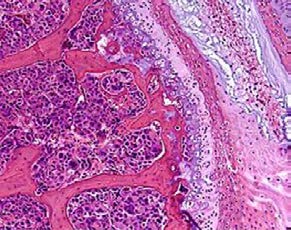
Paraffin section of mouse spine showing metastatic breast cancer, decaled section H&E Stain
Processing bone tissue requires highly specialized techniques, unique equipment, and technical expertise. Histological sections may be obtained on fresh frozen bones with mild decalcification, decalcified & paraffin-embedded bones, or on non-decalcified plastic-embedded bones. We also process all types of biomaterials, including engineering scaffolds and 3D co-cultured samples.
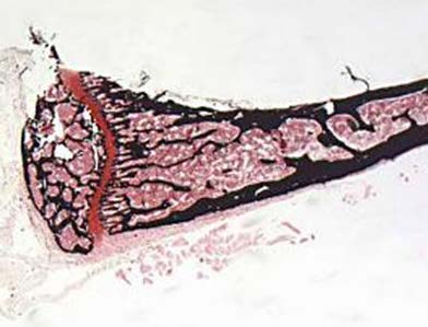
Plastic section of non-decaled mouse tibia, Von Kossa stain
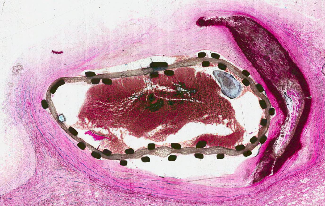
Ground section Human stented vessel with thrombus, methylene blue - basic fuchsin stain
The lab can provide service of producing ground sections on specimens which contain implants, by using the Exakt® cutting and grinding system.
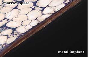
Ground section of Orthopedic implant
Ground section of impanted bone Methylene blue – Basic fuchsin
Histology procedures are performed on a broad range of experimental animal models including mouse, rat, rabbit, dog, monkey, and ferret. Other services include a wide array of special stains and immunohistochemistry, as well as histomorphometrical analysis of the histology sections.
Histomorphometry studies on bones may utilize standard bone histomorphometry (performed using the methods of Parfitt et. al.), or customized templates created for use with our Bioquant® Image Analysis Software. Histomorphometry can be used to estimate cell numbers, positively labeled cells, cell volume, or distance measurements. It can be used to measure the amount of protein present in a section by comparison with the integrated optical density of a known standard.
Four types of primary measurements are usually made—area, length (or perimeter), distance between point or lines, and number. These referents, such as tissue volume, bone volume, bone surface, and osteoid surface are used to derive other indices, such as trabecular number, trabecular thickness, and trabecular separation, as well as numbers of cell types per millimeter of bone surface.
Dynamic measurements of mineralized bones are made using unstained sections. Fluorescent measurements are made of single-labeled surface, double-labeled surface, and inter-label width. Applying the inter-label period, it is possible to calculate the Mineral Apposition Rate, as well as formation and resorption rates, and remodeling cycle duration.
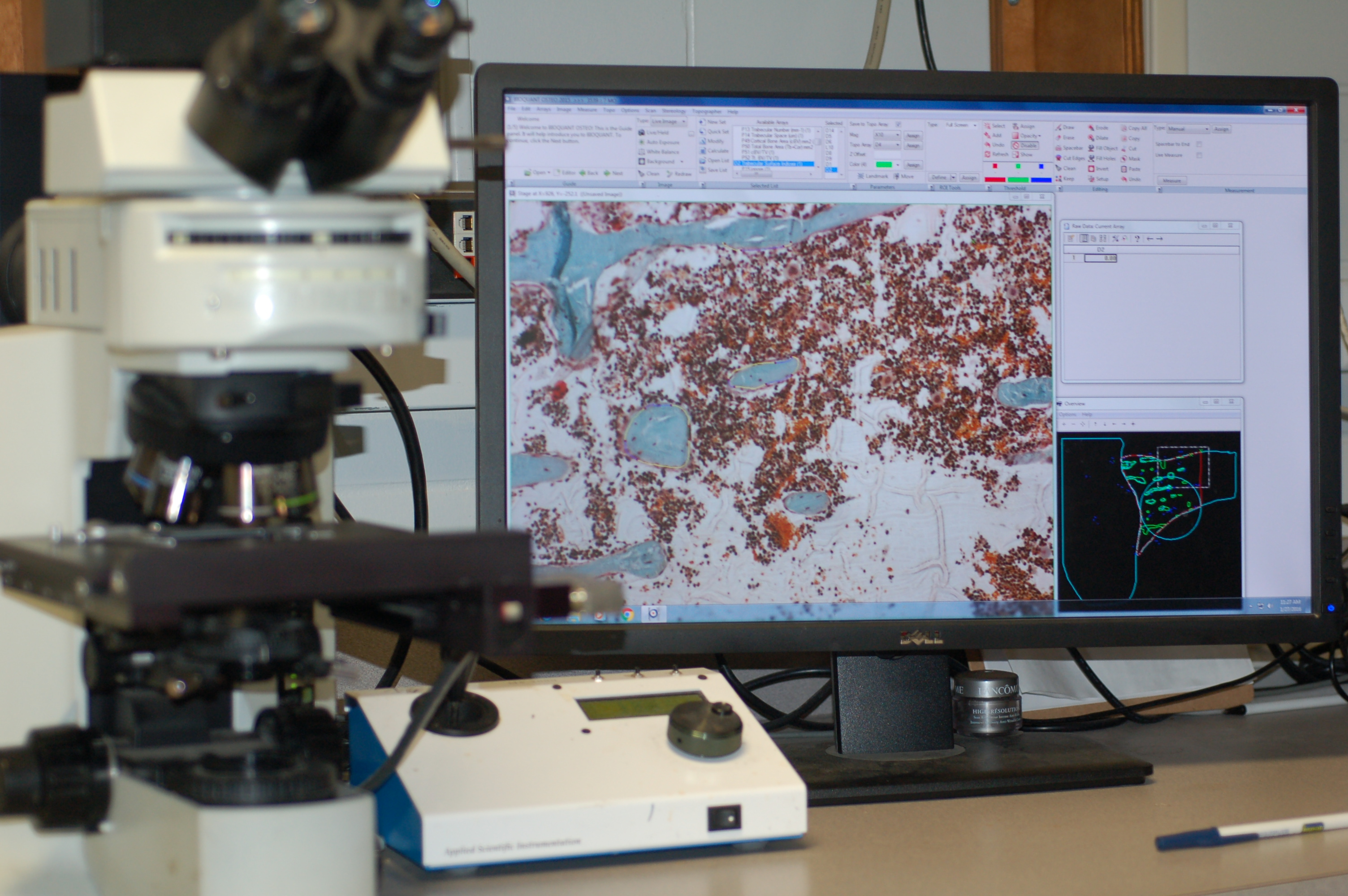
Bioquant System
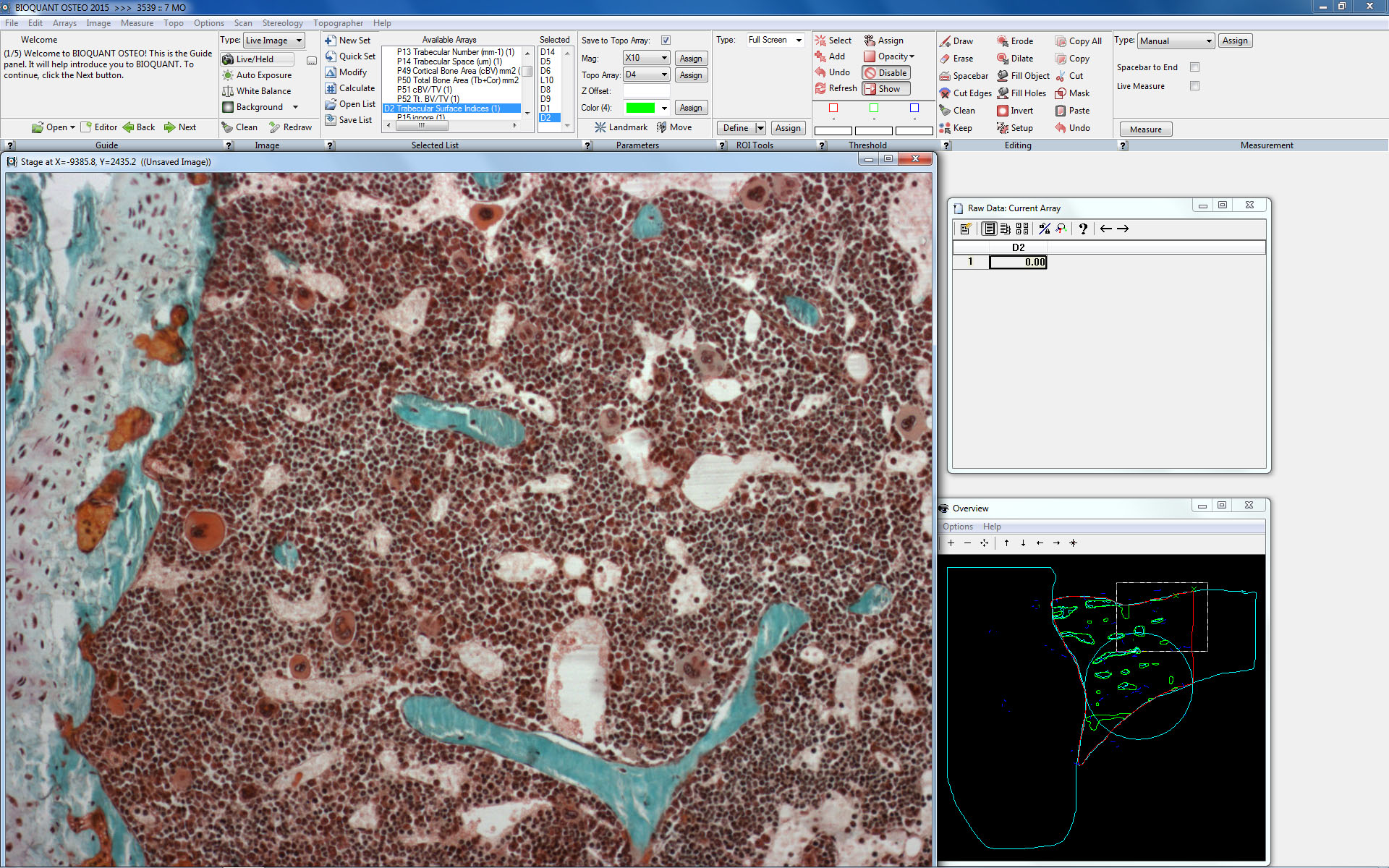
Bioquant Screen Image
Our laboratory has assisted researchers from departments such as Nutrition Science, Pathology, Microbiology, Gerontology, Hematology/Oncology, Biomedical Engineering, Orthopaedics, Neuropathology, Neuroscience, Rheumatology, Dental Biomaterials, Cell Biology and Physics.
We perform Immunohistochemistry and Immunofluorescent stains on a full range of samples (frozen or paraffin sections) with different species (mouse, rat, rabbit, dog , monkey and human, etc), & various type of tissues (soft tissues, xenografts, bones and teeth). We also provide consultation on antibody selection, tissue preparation and histomorphometric analysis.
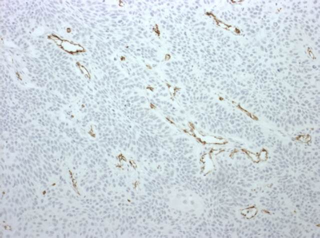
CD31 IHC stain on mouse xenograft paraffin section
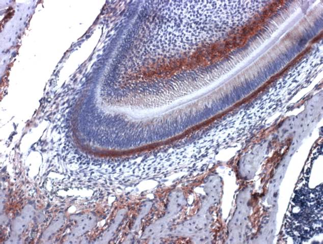
Alk Phos IHC stain on mouse tooth paraffin section
For more information and to obtain a copy of the “Order Form” contact the Laboratory Manager, Dezhi Wang, M.D., HTL, or Gene P. Siegal, M.D., Ph.D.