
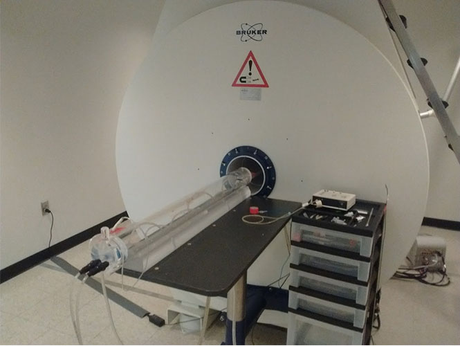
The Bruker 9.4T MRI (magnetic resonance imaging) scanner uses a strong cryogenic magnetic field, magnetic field gradients, and radio waves to provide high quality images of an animal. The small animal MRI can provide anatomical, quantitative and functional imaging. Different sequences, or acquisition settings, can be developed to generate various images for specialized analysis. Contrast agents can be used during MRI to improve any basic image, by enhancing the visibility of body structures.
This MRI provides high quality in vivo and ex vivo imaging of small animals including rats, mice, ferrets and tree shrews. The MRI system is equipped with isoflurane gas anesthesia and monitoring system enabling respiratory and ECG gating. Different radio frequency coils are available for imaging, including a volume coil, which can be used for full body imaging, and a surface coils, which can be used for imaging specific regions of interest. The small animal MRI offers unique opportunities for users to deliver preclinical data that can support translatable research.
Our MRI underwent a million-dollar upgrade in 2021 that greatly improved the reliability and capabilities of the MRI system and the quality of the images obtained. The new system is fully digital and has multiple receive channels for use with advanced phased array coils as well as a broadband RF amplifier for multi-nuclear studies of nuclei such as 13C, 19F, 31P, and 23Na with appropriate RF coils.