 |
Gorgas Case 2001-11 |
 |
|
The following case was seen in the outpatient department of the Regional Hospital in Iquitos, Perú. Iquitos, located on the banks of the Amazon is the largest city in the world (over 500,000 inhabitants) that is not reachable by road. The nearest road ends over 500 km away and all goods and people must come in by air or river.
We owe a large debt of gratitude to the following persons who were instrumental in getting these cases out each week during the 2001 courses. Carlos Seas, MD, Tropical Medicine Institute, Universidad Peruana Cayetano Heredia, Clinical Coordinator of the Gorgas Course for case selection and case histories; Tine Verdonk of the Institute of Tropical Medicine, Antwerp Belgium for digital photography; and Adam Plier, UAB Division of Geographic Medicine for web layout and coordination of electronic dissemination of the cases. These cases will be re-formatted to meet CME guidelines and will be available for online CME credit shortly. Please check back at www.gorgas.org or UAB CME in June of 2001. We look forward to providing more cases during the 2002 Gorgas Course. Please send any suggestions or comments for these cases to info@gorgas.org. |
|
The following patient was seen in the Outpatient Department of Cayetano Heredia National Hospital in Lima, Perú.
 History: Previously healthy
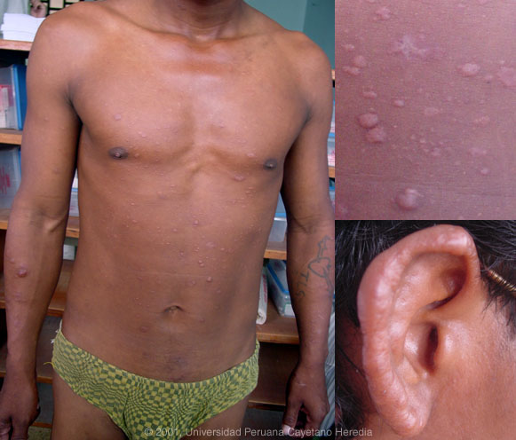
Epidemiology: Born in the high Andes but living in the jungle for the past 15 years. No exposures to animals or humans with infectious illness. No risk factors for Physical Examination: Positive findings restricted to nodular lesions shown in photograph. Lesions all over trunk and limbs as well as infiltrative lesions on the ears. No adenopathy, hepatosplenomegaly. CNS: normal motor function, normal cranial nerves, sensation normal to pin-prick. Labs/X-ray: Not performed except for skin examinations.
|
|
Diagnosis: Multibacillary leprosy. Lepromatous leprosy according to the Ridley-Jopling classification.
Discussion: Slit skin smears stained for acid-fast bacilli were 2+ from the left ear, 1+ from the left elbow, and 2+ from the left knee. With this classic clinical picture and with limited resources, this is all that is necessary for a definitive diagnosis and deep biopsy is not necessary. Slit skin smears are performed by making small (5mm length) slits in pinched skin (to avoid bleeding), the edges of which are scraped. The tissue fluid obtained is smeared on a clean slide and stained for AFB. Generally ear lobes, elbows, and knees are examined. The bacterial index ranges from zero (no bacillli in 100 oil-immersion fields) to 6+ (over 1000 bacilli in one field). The disease can be classified precisely in the immunologic sense using the traditional Ridley-Jopling classification. This is a spectrum of disease ranging from tuberculoid leprosy (TT) with no or few AFB in lesions and good cell mediated immunity to lepromatous leprosy (LL) with many AFB and poor cell-mediated immunity. However, the usual and more practical system is the WHO classification. For therapeutic purposes, it matters only whether the patient has paucibacillary or multibacillary disease. Paucibacillary leprosy is defined as five or fewer skin lesions with no bacilli on skin smears. Multibacillary cases have six or more lesions and may be skin-smear positive (approximately 10% of cases). Leprosy is a disease of skin and peripheral nerves. Although our patient had no neuropathy, but no examination of hot/cold sensation or light touch was documented, so subtle sensory abnormalities may well have been present. Leprosy is high on the diagnostic list in any patient with simultaneous skin lesions and sensory loss. Peripheral nerves such as the ulnar, median, common peroneal, posterior tibial, facial, and greater auricular are often palpably enlarged. In advanced neuropathy this often leads to motor deformities such as claw hand, footdrop, claw toes, and plantar insensitivity. Paucibacillary disease usually presents with small numbers of hypopigmented erythematous macules with absent sensation, well demarcated borders and some scaliness. Multibacillary disease is usually widespread at diagnosis with infiltrated, non-anesthetic papules or nodules with indistinct borders. The disease may be widespread without any distinct lesions. 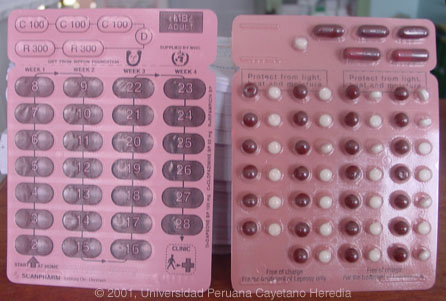
The standard WHO regimen for paucibacillary disease is 100mg Dapsone a day unsupervised and 600mg Rifampin once per month directly observed for 6 months. For multibacillary disease patients receive 100mg Dapsone and 50mg Clofazimine a day unsupervised and 600mg Rifampin and 300mg of Clofazimine directly observed once per month. A standard WHO multibacillary dose-pack is shown; the instructions in English must be clarified for all healthcare staff and patients. WHO now recommends only 1 year of therapy for multibacillary cases, but some would treat those with high bacterial indices (4 to 6+) for the previously recommended 2 years due to higher relapse rates. A myriad of treatment reactions can occur and a reference text should be consulted prior to initiation of therapy by anyone not familiar with these. |