 |
Gorgas Case 2001-07 |
 |
|
The following cases were seen on the ward of the Tropical Medicine Institute of Cayetano Heredia Hospital in Lima by the 2001 Gorgas Course participants.
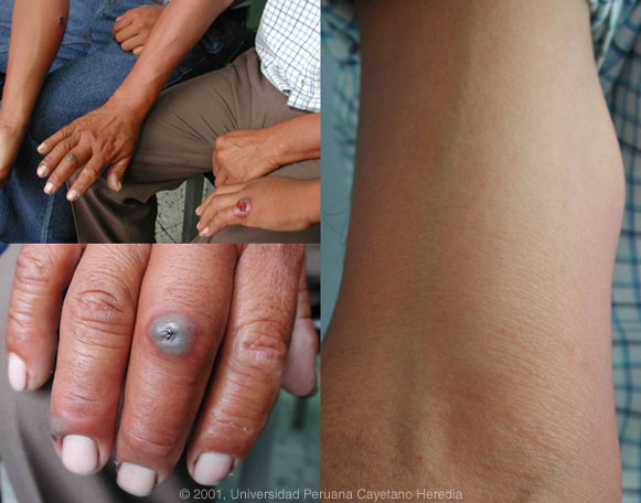
 History: Father (age 53) and 2 sons (ages 17 and 21) with hand and arm lesions shown in the photograph. 17 year-old developed a painless colorless blister on 5th finger of left hand, one day after exposure, which began to ulcerate 1 day later with edema and erythema of the entire hand [top left panel, hand on far right]. No fever, no adenopathy. Father developed painless papule on middle finger 48 hours after exposure [bottom left panel]. Painful adenopathy developed in the arm one day later [right panel]. No fever or systemic symptoms. 21 year-old developed blister on forearm 3 days after exposure with evolution to necrosis surrounded by erythema and edema [top left panel, arm on far left].
Epidemiology: All three are lifelong residents of Ica, an agricultural area surrounded by the coastal desert strip of Peru. 10 days prior to admission all three had participated in the necropsy of a cow that had died mysteriously. Two of the three recall cutting themselves. Physical Examination: Afebrile. No organomegaly. No findings other than that shown in photograph. Laboratory Examination: WBC normal. Normal hematocrit. Chest x-ray normal.
|
|
Diagnosis: Cutaneous anthrax.
Discussion: A gram stain of material from the still ulcerated lesion on the 17 year-old son showed highly typical large square-ended gram positive bacilli characteristic of Bacillus anthracis. Spores do not generally develop in vivo but only in soil, in hides, or culture. All three had received IV penicillin prior to transfer from Ica, so cultures taken in Lima were still negative at the time of being seen by Gorgas participants. Cutaneous anthrax constitutes over 95% of human infections with this organism and usually results from local inoculation into a wound or scratch. Incubation period is generally 1 to 7 days and our patients demonstrate a typical course. An initial papule develops a surrounding area of erythema and edema, and evolves into a vesicular or bullous lesion; sometimes, several satellite vesicles may develop. The fluid quickly becomes black from hemorrhage and then the papule ulcerates and develops a black central eschar with surrounding edema within several days. The lesions are generally painless but regional adenopathy (as in our patient) may be painful. Constitutional symptoms are unusual. Anthrax is a zoonosis which humans acquire predominantly from herbivores - cattle, sheep, horses, and goats - by direct contact with blood or other body fluids. Occurrence is worldwide in areas that support sporulation in the soil (at least 20°C). Soil, forage, and standing water are major reservoirs of spores by which animals acquire the infection. In industrialized countries, acquisition often occurs by contact with spores contaminating hair, wool or hides of infected animals. Other much less common routes of acquisition in humans (leading to more serious disease) are inhalational (spores from soil or environment) or gastrointestinal (ingestion of contaminated meat from sick animals). Human to human transmission is not thought to occur. Cutaneous anthrax is uniformly cured by penicillin G therapy. Tetracycline may be used in allergic patients. Oral therapy is likely adequate but 1 to 2 days of initial IV therapy is often used, especially if there is significant edema. Therapy sterilizes the lesions within 24 hours but does not modify the evolution of the lesions. Untreated the cutaneous infection may become septicemic with a mortality of up to 20%.
|