2001 Case #3 |
 |
|
| (Links to Other 2001 Cases are at bottom of this page) | ||
| The following case was seen in the outpatient clinic of the Tropical Medicine Institute of Cayetano Heredia Hospital in Lima by the 2001 Gorgas Course participants. |
 |
| History: One month history of 2 ulcerating skin lesions on the back of the hand at site of some subcutaneous nodules present since late October. One week after appearance of the initial 2 lesions, a 3rd lesion appeared above the elbow on the same arm. The lesions progressed to the present state despite local therapy and dicloxacillin. The lesions are non-painful, non-pruritic and have not been purulent at any time. No fever and the patient feels well. |
| Epidemiology: In late October this 32 yo policeman was dropped by helicopter, along with 7 other policemen, deep into the jungle on a mission he would not disclose the exact nature of. 3 days were spent walking and sleeping on the ground at night. Presently a Lima resident but has served off and on in jungle regions from |
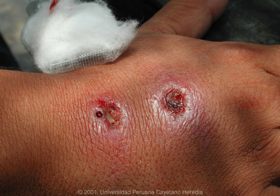
| Physical Examination: Afebrile. Hand lesions shown in photograph. Small subcutaneous nodules present in a lymphatic distribution up the forearm with a 3rd smaller similar appearing lesion just above the elbow. No lymphadenopathy. |
| Labs: Normal CBC and biochemistry. Aspirate and biopsy of lesions performed. |
2001 Case #3
|
|
|
| Diagnosis: Leishmaniasis due to presumed |
| Discussion: The major differential given the onset of the lesions on the hand and the lymphatic distribution would include leishmaniasis, sporotrichosis (endemic in Peru), atypical mycobacteria, and nocardiosis. In Peru leishmaniasis would be by far the most common. The painless nature of the ulcerative lesions, the characteristic heaped up borders, relatively clean bases are most indicative of leishmaniasis.
A needle aspirate of the heaped up border of the lesion revealed diagnostic intracellular amastigote forms of Leishmania on giemsa stain. PCR for speciation is pending. Of note, 3 of the other 7 accompanying policemen also have biopsy proven leishmaniasis with from 2 to 5 lesions. The ulcers are in varying sizes (0.5 to In South America it is important to distinguish Leishmania species that cause only cutaneous disease from the mucocutaneous species. Both cause initial skin ulcers but with mucocutaneous species, from months to years after treatment or healing of the skin ulcers, severe destructive recurrence may occur in the mucosal surfaces of the naso and oropharynx. Although speciation is pending in the Peruvian jungle, leishmaniasis is essentially exclusively due to Leishmania braziliensis, a species causing mucocutaneous disease. The patient was given therapy with pentavalent antimony (Glucantime) in a dose of
|