2001 Case #10 |
 |
|
| (Links to Other 2001 Cases are at bottom of this page) | ||
| The following case was seen in the outpatient department of the Tropical Medicine Institute of Cayetano Heredia Hospital in Lima by the 2001 Gorgas Course participants. |
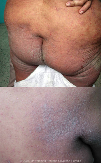 History: 53 yo female housewife with many years of non-specific skin symptoms. Over the previous 2 years this has progressed to a chronic dermatitis initially involving arms, shoulders, and axilla but spreading in recent months to involve buttocks and legs. Intensely pruritic, unable to sleep. No fever, no weight loss or systemic symptoms. No history of TB or exposure. Has had diagnoses of eczema, allergic dermatitis, and psoriasis, with intermittent treatment with antihistamines and local steroids. History: 53 yo female housewife with many years of non-specific skin symptoms. Over the previous 2 years this has progressed to a chronic dermatitis initially involving arms, shoulders, and axilla but spreading in recent months to involve buttocks and legs. Intensely pruritic, unable to sleep. No fever, no weight loss or systemic symptoms. No history of TB or exposure. Has had diagnoses of eczema, allergic dermatitis, and psoriasis, with intermittent treatment with antihistamines and local steroids.
Epidemiology: Lifelong resident of Lima except for 1 year in the jungle many years ago. Parents from Ayacucho and Abancay in the high Andes. No history of blood transfusions, IV or other illicit drugs; monogamous. Physical Examination: Afebrile. Skin lesions identical to those in photograph diffusely on body. No organomegaly or lymphadenopathy. Labs/X-ray: Hct 36. WBC |
2001 Case #10
|
|
|
| Diagnosis: Norwegian (crusted) scabies secondary to |
| Discussion: Skin scrapings viewed by direct microscopy disclosed The crusted diffuse highly pruritic skin lesions in this patient are very characteristic for Norwegian scabies. This condition is well described in many immunocompromising conditions including HIV infection, malignancy, and immunosuppressive therapy. The skin is hyperinfested with thousands of mites, so it is easily diagnosable with simple scrapings. The lesions are highly infectious and present a serious nosocomial risk. At the Tropical Medicine Institute in Lima approximately Other conditions associated with The prevalence of Norwegian scabies is not responsive to normal topical agents such as benzyl benzoate or permethrin. Our patient was treated with ivermectin |