2002 Case #5 |
 |
|
| During Week 5 of the course, the annual field trip to Cusco in the Andean highlands took place. Cusco (elevation 3400m) is the oldest continuously inhabited city in the Americas. The patient was seen in the Hospital Regional de Cusco. We thank gastroenterologist Dr. Ernesto Cazorla for permission to use his endoscopic images. |
 History: 72 yo with 3 day history of acute onset severe epigastric pain admitted initially to the surgical service. No fever. No significant past medical history. No chronic or recent medications. History: 72 yo with 3 day history of acute onset severe epigastric pain admitted initially to the surgical service. No fever. No significant past medical history. No chronic or recent medications.
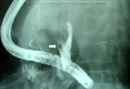
Epidemiology: Impoverished urban dweller. No TB exposure. No ethanol history. No ill contacts. Physical Examination: Afebrile. Acutely tender abdomen predominantly in the epigastric area but without peritoneal signs. Normal rectal exam. Labs/X-ray: Hematocrit 42. WBC 12,200 with 70 segs, 10 bands, 5 monos, 13, lymphs and 2 eos. Amylase 500 (N=40). Bilirubin 1.8, direct predominance. SGOT 2X normal. Normal BUN and creatinine. Abdominal CT was read as compatible with acute resolving pancreatitis. An ERCP was performed and after papillotomy dye injected into the common bile duct; the arrow on the image demonstrates the relevant filling defect.
|
2002 Case #5
|
|
|
| (Links to Other 2002 Cases are at bottom of this page) | ||
| Diagnosis: Pancreatitis and acute biliary obstruction due to Ascaris lumbricoides. |
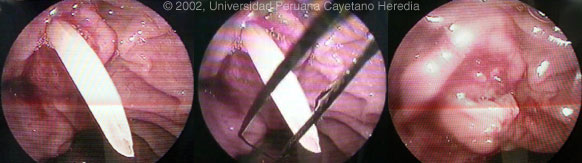
| Discussion: The lengthy dye-free filling defect (shown by the arrow in previous image; also shown again at bottom of this page) obstructing one of the main hepatic ducts, and which had extended back through the papilla of Vater, corresponds to the shape of an adult Ascaris lumbricoides that had aberrantly migrated into the biliary system from its normal home in the intestinal lumen. The images below show direct endoscopic visualization of the tail end of the adult worm sticking out of the common bile duct after the endoscopic papillotomy. In real time the worm can be seen moving back and forth as it migrates up the biliary tree. Through a wider papillotomy the tail end of the worm was grabbed with the forceps as shown in the digital image and physically removed, thus obviating the need for laparotomy in this case. The final image shows the common bile duct opening after the worm was removed.
 Ascaris lumbricoides is the commonest of the intestinal nematodes of humans, with very few unaffected countries, and affecting approximately one-quarter of the world's population. Each adult female produces up to 200,000 eggs per day which once passed in the feces can remain infectious in the environment for decades. Infection is caused by oral ingestion of fecally contaminated material. As can be seen from the images these are the largest of the human intestinal nematodes and when an adult is incidentally passed in the feces by a patient it rarely goes unnoticed. The overall complication rate is low, of the order of less than 1 in 1,000. However, the high prevalence of ascaris infection makes these manifestations a not uncommonly seen clinical syndrome in highly endemic impoverished areas. Intestinal obstruction, which is often fatal if treatment is not readily available, due to a bolus of large numbers of adult worms is most common, especially in children under the age of 10 (who have smaller intestinal lumens). Less common are obstructive lesions due to migratory adult worms which may block the pancreatic duct, the common bile duct, a main hepatic duct, or end up in the liver parenchyma and cause an abscess. In the intestine, worms may migrate and obstruct the appendix or the lumen of a diverticulum. Rupture of a diverticulum or a major bile duct rarely occurs. The totality of precipitating factors for migratory ascaris are not well characterized but certainly recent anthelmintic therapy or general anesthesia (which may partially relax worm musculature) are associated in some cases. Eosinophilia is usually seen only where there is actual tissue invasion by any one of the helminthic parasites. Adult ascaris live in the lumena of the gastrointestinal tract (intestine, biliary tree uncommonly) and do not invade tissue. Thus, the lack of eosinophilia in this case is not surprising. However, ascariasis can transiently be associated with eosinophilia in the early phase after initial infection. Immature larvae migrate through the lung and may cause a transient eosinophilia due to the tissue invasive nature of the brief phase of the life cycle. In cases of biliary obstruction, endoscopic removal of the adult worm is relatively simple and is now considered by us to be the treatment of choice. Much literature and many textbooks still recommend conservative therapy with nasogastric suction and older anthelmintic drugs such as piperazine which relax but don't kill the ascaris. This potentially allows the worm to be passed with the normal motility of the biliary tree. While this approach may often be successful, the recommendations come from centers where endoscopy was not readily available and the only other option would have been laparotomy, which may still be necessary if conservative therapy fails. This latter approach results in prolonged hospitalizations and potentially prolonged patient discomfort. There are currently large numbers of immigrants and refugees living in industrialized countries and ascariasis is a highly prevalent infection. The diagnosis should be considered in any case of pancreatitis or biliary obstruction in this patient population.
|