Explore UAB

University of Alabama at Birmingham

Gorgas Case 2021-04

Universidad Peruana Cayetano Heredia

The following patient was seen as an outpatient at a private clinic in Lima.

Image for Case 2021-04

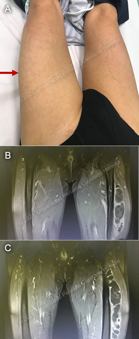
History: A 75-year-old male patient was transferred from a hospital in the highlands of the country to a private clinic in Lima for further evaluation of progressive swelling of the left thigh that ensued over the last 5 months. The lesion was painless, it was not associated with systemic symptoms or physical impairment, and was not related to previous trauma, he also denied similar lesions in other parts of the body or a previous febrile episode. He remembered to have had a similar but smaller lesion in the left thigh in 2016 that was treated with unknown antibiotics with full resolution.

Epidemiology: Born and lives in Huancayo, a city in the highlands of Peru. He works as a farmer and was in contact with dogs, cats, guinea pigs and pigs throughout his life. Denies any recent travel. Denies contact with TB patients.

Physical Examination: Afebrile with normal vital signs, the patient appears in no distress. There was a non-tender soft swelling of the left thigh with no inflammatory signs on the overlying skin. (Image A). Chest and cardiovascular examinations were normal. No organomegaly. Normal neurologic examination.

Imaging studies: Chest x-ray was normal. MRI of the left leg is shown in Images B and C. The abdominal ultrasound was reported as normal.

Laboratory Examination: Hb: 13.7 g/dL; Hct. 40%; WBC 7 650 (neutrophils: 60%, eosinophils: 0, lymphocytes: 35%); Platelets: 236 000. Gluc: 108 mg/dL, Creat: 0.9 mg/dL, AST 33 U/L, ALT 27 U/L, albumin 3.9 g/dL.


UPCH Case Editors: Carlos Seas, Course  Director / Paloma Carcamo, Associate Coordinator

UAB Case Editors: David O. Freedman, Course Director Emeritus / German Henostroza, Course Director

{slide=CLICK HERE FOR DIAGNOSIS & DISCUSSION OF THE CASE ABOVE

Diagnosis: Primary skeletal muscle hydatidosis caused by Echinococcus granulosus

Images for Case 2021-04
image overlay

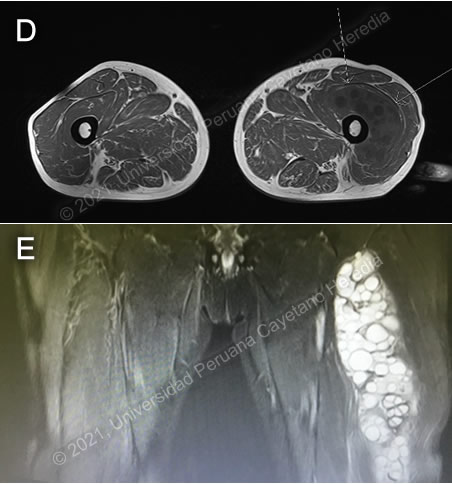
Discussion: The serum Western-Blot for E. granulosus infection was positive. The MRI of the left leg shows (T1 sequence with contrast) the presence of a large hydatid cyst with daughter cysts inside located in the left thigh (Images B and C). Additional images at the T1 sequence without contrast reveals that a large cyst is located in the intermediate vast of the quadriceps femoris muscle, the T2 weighted sequence revealed high signal intensity characteristic of the liquid component of the cysts (Images D,E). On further questioning, he remembered that at the age of 14 years a small cyst was identified in his right upper lung, medical treatment was offered but he does not remember further clinical or therapeutic details.

Primary skeletal muscle hydatidosis is a very rare complication of cystic hydatidosis accounting for less than 5% of all cases, and results from hematogenous dissemination of the larval stage (oncospheres). Concomitant liver and lung cysts can be observed as well. Trapping of the cysts in the capillary vessels of the liver and lungs may explain the rarity of this and other locations. The typical cystic formation may not occur in the muscles as a result of mechanical movement of the muscles and high lactic acid concentration [Can J Surg 1974;17:232]. Secondary muscle hydatidosis, or recurrent disease, results from spilling of the cystic content during a surgical procedure specially if post-surgical treatment was inadequate or not offered, or due to traumatic rupture of the cyst with subsequent spread of its content [PLoS Negl Trop Dis 5(1): e840]. The most common muscles involved are the thigh, pelvic and paravertebral muscles, including the psoas muscles [Clin Infect Dis 2001;32:e65]. The clinical presentation is related to the dimensions of the cyst, and includes pain, deformity of the area involved, limitations to perform physical activity, vascular insufficiency due to compression and complications such as anaphylactic reactions due to the partial or total rupture and superinfections. In rare cases, bone involvement may occur with osteolytic lesions mimicking neoplastic or bacterial involvement. Diagnosis may be difficult as the clinical picture and the images mimic other conditions such as cancer or pyogenic infections. Serology may be negative as well. High index of suspicion and proper reading of images including ultrasound, CT Scan or MRI are very useful. Ultrasound is the imaging of first choice, when non diagnostic, a CT-Scan may help showing daughter cysts and increased density of the hydatid membrane [Parasitol Int 2008;57:8]. Findings at the MRI include at T1 diverse patterns of signal intensity and at T2 the liquid content of the cyst is revealed, but these findings are non-specific [Clin Infect Dis 2001;32:e65; Int J Sur Case Reports 2018;51:379].

Human hydatid disease secondary to Echinococcus granulosus is caused by the larval form of this dog tapeworm. Humans ingest the tapeworm eggs in environments contaminated by canine feces and become accidental intermediate hosts. This patient had ongoing exposure to dogs during his entire life. Sheep are the normal intermediate hosts. In general, disease is diagnosed in adulthood as larval cysts expand slowly over years or decades, becoming symptomatic as they impinge on other structures by virtue of their size. The cysts contain hundreds of viable protoscoleces capable of becoming adult tapeworms upon ingestion by a definitive host such as the dog. The internal germinal membrane lining the cyst produces new protoscoleces on an ongoing basis and may also produce internal daughter cysts. Each protoscolex is capable of becoming a new cyst should the original cyst rupture or be ruptured. Cystic hydatid disease due to E. granulosus is common in sheep and cattle raising areas worldwide. Most primary infections involve a single cyst. In adults, 65% of solitary cysts are found in liver, 25% in lung and the rest in a wide variety of other organs including kidney, spleen (see case #7, 2009), heart, bone, muscles and brain (see case #5-2014). This is the first case of a well-documented skeletal muscle hydatidosis presented as a case of the week since we started posting cases in 2001.

Differential diagnosis of swelling of the thighs includes malignancy, hematoma, osteomyelitis and pyogenic myositis. Diagnosing malignancies requires a tissue sample. Pyogenic myositis in the tropics is more commonly seen in children (tropical pyomyositis, see case #9-2002), aspiration of the abscess reveals purulent material, Staphylococcus aureus is the most common pathogen involved (95% in the tropics), patients are usually febrile and with marked tenderness. Chronic osteomyelitis with swelling of soft tissues and drainage of purulent material through sinus tracts is easily recognized in a plain x-ray film.

The treatment of choice for skeletal muscle hydatidosis is surgical removal of the cyst. The WHO recommends pre- and post-surgical treatment with albendazole. Duration of pre-surgical treatment is not well defined, courses as long as 3 months resulted in 94% elimination of viable cysts [PLoS Negl Trop Dis 5(1): e840]. Post-surgical treatment for one month with albendazoles is recommended by WHO. For patients in whom the surgical resection is not possible, or in those with recurrent disease after surgery the PAIR procedure (puncture, aspiration, injection and re-aspiration) plus antiparasitic treatment may be an option, but there is no experience with this procedure in muscle hydatidosis [PLoS Negl Trop Dis 5(1): e840]. Our patient was scheduled for surgical resection after receiving a course of albendazole plus praziquantel for one month but he acquired COVID-19 and was lost to follow-up.

{/slide}