Explore UAB
University of Alabama at Birmingham

Gorgas Case 2023-07

Universidad Peruana Cayetano Heredia
The following patient was seen as an inpatient in the Tropical Medicine ward of Cayetano Heredia Hospital in Lima by the 2023 Gorgas Course participants.
Image for Case 2023-07

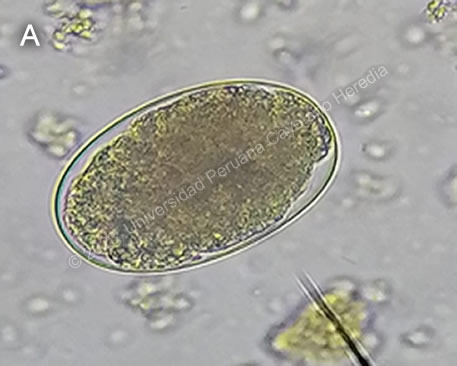
History: A 22-year-old previously healthy male patient presented with a 3-month history starting with mild dyspnea on exertion and fatigue. The dyspnea and fatigue worsened in the following weeks; two weeks before admission he stopped working due to the dyspnea. Ten days before admission, he had an episode of mesogastric pain which subsided using natural medicine. On the day of admission, he had nausea and one episode of vomiting, accompanied with fever, dizziness, and headache.

Epidemiology: Born and lives in Yurimaguas, Loreto, in the Amazon Basin. Traveled from Loreto to Lima three weeks before admission. Works as a carpenter. Drinks alcohol once a month, smokes four cigarettes per day, has used cocaine paste in the past.

Past medical history: No other illnesses, no past surgeries.

Physical Examination:
 BP 100/80 mmHg, HR 109 bpm, RR 20 rpm, Sat 98% (FiO2 0.21). Skin: Marked pallor. No skin lesions. Rest non-contributory.

Laboratory: Hemoglobin 2.6 mg/dL (MCV 80.3, MCH 23.6), hematocrit 10, WBC 16 190 (neutrophils 10 900, eosinophils 2 580, basophils 50, monocytes 750, lymphocytes 2 280), platelets 300 000. Ferritin 3 ng/mL (21.8-274.6), vitamin B12 198 pg/mL (normal 187-883), folic acid 4.6 ng/mL (normal 3.1-20.5). PT 13, aPTT 30, INR 0.98. Glucose 77, urea 23, creatinine 0.83, Na 137, K 4.8, Cl 95. AST 30, ALT 28, LDH 166, Alk Phos 122, GGT 22. HIV, RPR HBsAg non-reactive. Thick and thin smear were negative. AFB in sputum was negative x3. PPD was 0mm. Stool O&P is shown in Image A.

Imaging: Chest X ray and abdominal ultrasound showed no abnormalities.


UPCH Case Editors: Carlos Seas, Course  Director / Paloma Carcamo, Associate Coordinator

UAB Case Editors: David O. Freedman, Course Director Emeritus / German Henostroza, Course Director

{slide=CLICK HERE FOR DIAGNOSIS & DISCUSSION OF THE CASE ABOVE

Diagnosis: Severe anemia secondary to hookworm infection.

Images for Case 2023-07
image overlay

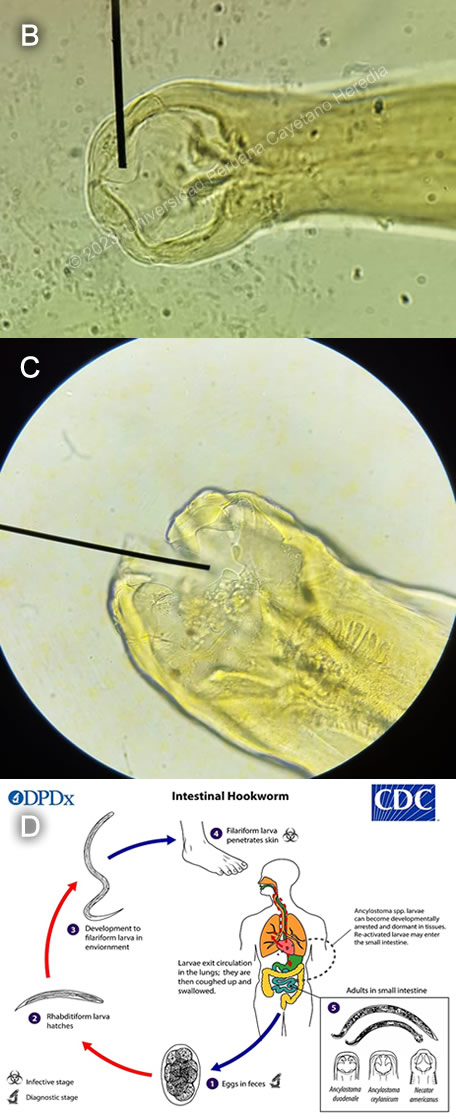
Discussion: The stool O&P exam revealed hookworm eggs, Baermann was negative for Strongyloides. It is not possible to differentiate Necator americanus from Ancylostoma duodenale from examination of the eggs. The sample was cultured, and examination of the adult roundworm revealed the etiologic agent for our patient’s anemia to be Necator americanus (Image B), with characteristic ventral plates in the buccal capsule in contrast to the teeth that can be seen in Ancylostoma duodenale (Image C). Upon further questioning, the patient confirmed he regularly walked barefoot in the jungle when searching for timber. This patient likely had a very chronic infection with hookworms with a slow decline in hemoglobin levels over years, which only became symptomatic in the months before admission.

Hookworm infection is transmitted through contact with soil contaminated with third-stage larvae, which penetrate the skin causing a localized, pruritic, erythematous-papular rash known as “ground itch”. Larvae then enter the bloodstream until they reach the heart, whereupon they migrate to the alveolar spaces through the capillaries, are coughed up and swallowed and finally reach the small intestine, where they mature, reproduce, and produce eggs. The eggs are passed in the stool and into the environment, where they hatch and mature until they are ready to perpetuate the cycle1 (See Image D, copyright CDC2).

Chronic infection with hookworms presents clinically as iron-deficiency anemia due to blood loss. Parasites use their buccal apparatus to attach to the intestinal wall, breaking capillaries and arterioles to feed. Blood loss is both due to the parasites feeding on blood directly and to their secretion of anticoagulant substances. Infection with A. duodenale seems to cause more blood loss than infection with N. americanus, but both hookworms can cause significant anemia, particularly after the host’s iron stores have been depleted. Hookworm infection usually causes eosinophilia during larval migration through the lungs (Loeffler’s syndrome); in the case of our patient the eosinophilia is most likely due to repeated infections caused by persistent exposure.

There are many other causes of anemia in the tropics, including other parasitic infections such as malaria or diphyllobothriasis, though the former will cause acute hemolytic anemia and the latter B12 deficiency anemia. Other helminths such as Trichuris trichiura or Ascaris lumbricoides may cause gastrointestinal complaints but are not typically associated with anemia. A diagnosis of hookworm infection can be established with a stool exam showing typical eggs. The eggs of the two species of hookworms are indistinguishable, but they can be differentiated using PCR or culturing the larvae. Antiparasitic treatment for hookworms should be given with albendazole, which has better egg reduction rates than mebendazole. However, either drug can be given for mass treatment public health interventions, as mebendazole has better egg reduction rates for treatment of T. trichiura, and both are effective against A. lumbricoides. Treatment with albendazole in the setting of targeted therapy or mass treatment campaigns is given as a single 400mg dose; mebendazole is also usually administered as a single 500mg dose during campaigns, but can also be given as a twice-daily 100mg dose for three days. Our patient received treatment with a single dose of albendazole, blood transfusions, and was discharged after marked clinical improvement.

References:
1. Hotez PJ, Brooker S, Bethony JM, Bottazzi ME, Loukas A, Xiao S. Hookworm Infection. New England Journal of Medicine. 2004;351(8):799-807. doi:10.1056/NEJMra032492
2. Prevention CC for DC and. CDC - Hookworm - Biology. Published September 17, 2019. Accessed March 15, 2023. https://www.cdc.gov/parasites/hookworm/biology.html


{/slide}