 |
Gorgas Case 2026-5 |
|
The following patient was seen on the inpatient ward of Cayetano Heredia Hospital in Lima by the 2026 Gorgas Course participants.
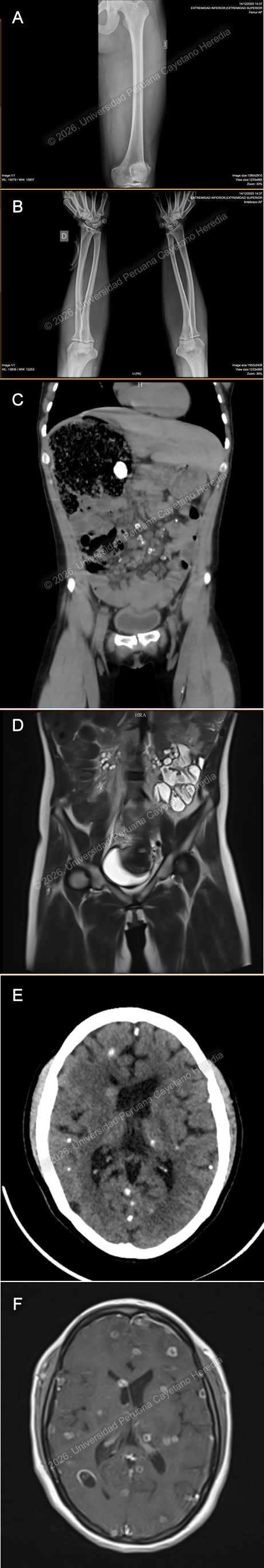 History: A 25-year-old woman from the highlands of Huancavelica presented with a 4-year history of gradually worsening abdominal pain. Initially, she experienced mild, diffuse upper abdominal pain after meals, which was later accompanied by nausea, decreased appetite, and postprandial vomiting (≥2 episodes daily). Nine days before admission, her condition worsened with increased fatigue and general malaise. Due to persistent symptoms, she sought hospital care and was admitted for further evaluation and management. On review of systems, she describes mild to moderate global headaches and subjective fevers but denies visual disturbances or a history of seizures. Epidemiology: The patient was born with an imperforate anus and underwent stoma surgery at 1 year old at Cayetano Heredia Hospital. She is originally from and currently lives in Izcuchaca, Huancavelica, a rural highland area in Peru, at 2,900 meters above sea level. She sells food on the streets and has incomplete primary education. She lives with relatives in a household without access to potable water, and reports multiple exposures to farm animals (hens, cows, guinea pigs, pigs) and currently owns sheep. She denies exposure to vectors, travel history, and sick contacts. Physical examination: HR: 81 bpm, RR: 20 breaths per minute, T: 36.5°C, BP: 100/70, SpO2: 97% on room air. On abdominal examination, a stoma was noted in the epigastric region, with tenderness on palpation of the upper abdomen. The rest of the physical exam was unremarkable. An imperforate anus was also present. Laboratory: Initial laboratory tests showed a normal hemoglobin of 12.2 and leukocytes of 4400; with 53.4% neutrophils, 36.6% lymphocytes, 0.3% basophils, 4.9% monocytes, and 4.8% eosinophils. Platelet count was 221,000. Liver function tests revealed AST 79 U/L (normal value 8 to 48 U/L) and ALT 65 U/L (Normal range 7 to 55 U/L). Alkaline phosphatase of 25 U/L (normal range 30 to 130 U/L), and GGT of 184 (normal range 5 to 40 U/L). Total bilirubin was 0.3, with direct bilirubin 0.2. Renal function was within normal limits, with urea 27 mg/dL and creatinine 0.7 mg/dL. Electrolytes were also normal, including sodium 137 mEq/L, potassium 4.63 mEq/L, and chloride 102 mEq/L. Albumin was 4.4 g/dL. HIV and RPR were negative. Imaging: Upper and lower limb X-rays revealed multiple calcifications in the muscles (Images A, B), Abdominal CT showed hepatic nodules, ileocolic lymphadenopathy, and a distended ascendant colon with cystic lesions in the mesenteric adipose tissue (Image C), multiple cystic lesions are also evidenced in the pelvic MRI (Image D). Neuroimaging revealed multiple intracranial cystic and calcified lesions on head CT (Image E), and brain MRI confirmed multiple intraparenchymal cysts with ventricular involvement (Image F). UPCH Case Editors: Carlos Seas, Course Director / Paola Nakazaki, Associate Coordinator |
|
 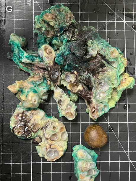 Discussion: A serum Western Blot assay was positive for cysticercosis. Cysticercosis is a neglected tropical disease and a major global public health issue, most common in developing countries, especially in regions of Asia, sub-Saharan Africa, and Latin America (1). In Peru, the highest prevalence occurs in the highlands, particularly in Cuzco, Huancayo, and Andahuaylas; on the northern coast in Tumbes, and in some areas of the high jungle (2). The parasite has a two-host life cycle involving a definitive host and an intermediate host. Humans are the only definitive hosts for the adult tapeworm and can also serve as accidental intermediate hosts. Pigs are the typical intermediate hosts in endemic areas, harboring the larval form in their tissues (3). Cysticercosis is acquired through the fecal-oral route after the ingestion of Taenia solium eggs in contaminated food or water. After ingestion, the eggs hatch into oncospheres, which penetrate the intestinal wall and disseminate hematogenously to various tissues, where they develop into cysticerci and can localize in muscle, subcutaneous tissue, eyes, and particularly the central nervous system (1,3). The infection can stay asymptomatic throughout life, especially in endemic areas, or show a range of clinical signs depending on the location, size, number, and stage of the lesions (4). These features can also influence prognosis and treatment options. Diagnosis mainly relies on imaging studies, supported by serologic tests such as ELISA and Western Blot (5). Our patient presents with disseminated cysticercosis involving soft tissues, the intestine, and the brain. In this patient, NCC was characterized by inactive calcified lesions, active cystic lesions, and intraventricular cystic lesions. Clinical presentation of disseminated cysticercosis is rare and affects the brain, muscles, subcutaneous tissue, eyes, and, sometimes, organs such as the liver and heart. It is diagnosed when cysticerci are found in at least two separate organ systems, confirmed by imaging or histopathology. It has been associated with risk factors including residence in highly endemic areas, and altered immune response (7,8). Given the uncommon nature of this presentation, clear management guidelines have not yet been established (9). Our patient underwent a right colectomy with extended ileal resection, jejunum–transverse anastomosis, removal of mesenteric adipose tissue, and colostomy revision. Histopathological examination of the surgical specimen demonstrated segments of small and large intestine with multiple cystic lesions involving the submucosa of the small intestine and the mesentery, some of which were calcified. Parasitic structures consistent with cysticerci were identified in the mesenteric adipose tissue (Image G). She is currently awaiting endoscopic removal of the intraventricular cyst, which will be followed by antiparasitic treatment. References |