 |
Gorgas Case 2003-01 |
 |
|
The Gorgas Courses in Clinical Tropical Medicine are given each year at this time at the Tropical Medicine Institute at Cayetano Heredia University in Lima, Perú. For the third consecutive year, we are pleased to post interesting cases seen by the participants during the courses. New cases will be posted every Monday for the duration of each course. Each case includes a brief history and a digital image pertinent to the case, followed by a second page with the diagnosis and brief discussion. Currently, the 2-week Gorgas Expert Course is running and will be immediately followed by the 9-week basic Diploma course, so that 11 weekly cases are anticipated this year.
|
|
The following patient was seen in the Outpatient Clinic of the Tropical Medicine Institute.
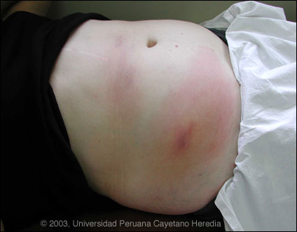
 History: 57 year-old female with 10-day history of highly pruritic rash which began as a painful violaceous nodule in the mid upper abdominal region with surrounding erythema and extreme warmth. Her physician started ciprofloxacin, which was switched to Augmentin after 5 days. As the inital lesion was resolving, the pruritus and erythema tracked down to the right lower quadrant with development of a second painful nodule. At the time of presentation the patient felt the pruritis continuing to spread further towards the right flank. No headache, fever, chills, or other abdominal or gastrointestinal symptoms.
Epidemiology: From an upper class district of Lima. Onset of symptoms while returning from a 3-week holiday visit with family in Texas. Two days prior to the onset of illness a meal eaten over the border in Mexico included shrimp, prawns, and perhaps other fish. In Lima, the patient eats Ceviche once to twice per week in restaurants. A month prior to illness onset she had eaten at a new restaurant in Lima that had been highly recommended. No exposures to animals or other ill people. No recent other travel. No beach visits since summer last year. Physical Examination: Afebrile. Two resolving, presently non-tender, violaceous nodules with large surrounding erythematous reactions as shown in the Image. No organomegaly or lymphadenopathy. Normal neurologic examination. Laboratory Examination: Hematocrit 43. WBC 7.6, 53 neutrophils, 32 lymphocytes, 6 lymphocytes, and 8 eosinophils (608 total eosinophils). AST 27, ALT 32.
|
|
Diagnosis: Gnathostomiasis.
Discussion: Gnathostomiasis, a disease that is most highly endemic in and associated with Southeast Asian countries such as Thailand, Vietnam, and China, is very much an emerging disease of travelers especially in Latin America. First reported in Mexico in 1970 now over 1000 cases, mostly from the western part of the country and including well-visited resort areas such as Acapulco have been reported in the past decade. Significant numbers of cases have now been reported from Ecuador and in Perú we now have experience with several dozen cases in the past 5 years. Human cases are caused by Gnathostoma spinigerum. Human infection is almost always acquired as a result of ingestion of immature larvae in undercooked freshwater fish. The adult worms live in tumors in the stomachs of dogs and cats. Eggs are passed via the feces and and resulting larvae ingested by minute copepods in freshwater which are in turn ingested by a an indiscriminate range of fish, frogs, as well as reptiles. When accidently ingested by humans the immature larvae cannot become mature adults in the intestine but rather can migrate widely through subcutanous as well as deeper tissues. Our patient demonstrates the most frequent clinical manifestation, that of inflammatory and mildly hemorrhagic necrosis along the track of the migrating larva. The presentation is almost always of transient subcutaneous nodular lesions that appear in sequence along the track of the larvae. This pattern is usually different from the typical well-demarcated and thin red linear serpentine lesions of creeping eruption (cutaneous larva migrans) due to Ancylostoma species of animals or the very rare instances when Fasciola larvae migrate subcutaneously. In endemic areas of Africa the migratory Calabar swellings of Loa loa infection and in China migratory Paragonimus larvae would be a consideration. Subcutaneous nodules can be present with onchocerciasis, cysticercosis, or sparganosis but don't present a migratory pattern. Less frequently in Gnathostoma infection, the larvae can migrate to vital organs, including the CNS (eosinophilic meningitis), lungs, and eyes (with severe conjunctival edema), and the disease rarely can be fatal. Diagnosis may be made by direct visualization of the larvae in biopsy material. The yield of biopsy is sub-optimal due to the ongoing movement of the migratory larvae and is often unecessary if the patient has a classical presentation. When therapy is initiated dying larvae may migrate closer to the surface to a fixed position so newly dead larvae may be easier to isolate. Serology is not widely available. An ELISA developed by Dr. Jitra at Mahidol University in Bangkok is reported to have close to 100% sensitivity and specificity for G. spinigerum. 17 of our local cases have been confirmed in this way and serological confirmation of the present case is pending. Ceviche is a fish dish where pieces of fish are marinated in lime juice and eaten with raw vegetables. Lime juice is ineffective in killing the larvae of Gnathostoma. In Mexico, ceviche made from freshwater fish such as tilapia is common. Our cases have typically been associated with ceviche ingestion, however, ceviche in Lima is never made with river fish. No systematic study of local fish populations has been undertaken yet in Perú. Nevertheless, it is interesting to note that all our cases have been from the high socio-economic classes in Lima where ceviche made from corvina (sea bass) and lenguado (flounder) is most commonly ingested. Corvina spends part of its life cycle near the mouth of freshwater estuaries. In Guayaquil, Ecuador, Gnathostoma has been definitively isolated from local corvina (Dr. José Ollagué, Personal Communication). We cannot implicate the meal taken by this patient on the Mexico-Texas border as that ingestion was mostly shrimp, not fish, and the symptoms began less than 48 hours after that. This is a shorter incubation than would be expected with gnathostomiasis. Both Albendazole 400-600 mg/day for 2-3 weeks and ivermectin 150-200 ug/kg/day for 1-3 days have been reported to have cure rates in the 95% range. Patients that don't respond to one agent often respond easily to the other or to a second course of the same agent. Relapses do occur and usally respond well to re-treatment although most would use the other agent, if available, during a relapse.
|