Focus on some of the Ocular Phenotyping & Molecular Analysis Module Equipment
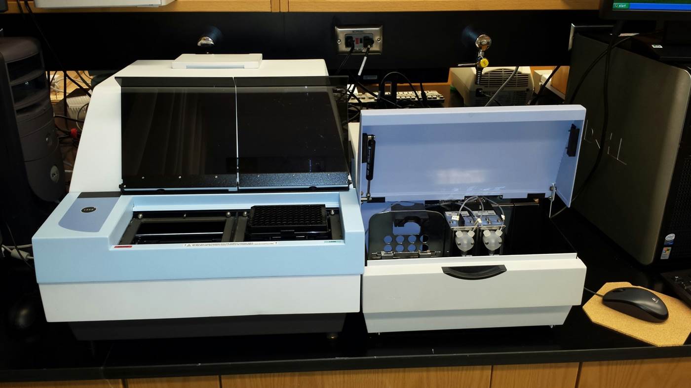
- Fluorescence (top and bottom; 340-850 nm)
- Luminescence (glow, flash, dual)
- Photometry (340-800 nm and UV 260/280)
- Time-resolved fluorometry (dual window; dual emission)
- Fluorescence polarization (400-850 nm)
2 Leica CM3050 S Cryostats
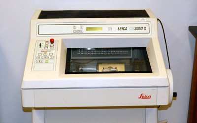
For cryosections of fixed and unfixed tissue specimens, capable of producing step or motorized sectioning for high-quality sections of consistent thickness, that allows:
- Preservation of enzyme activity for enzyme histochemistry and antigenicity for immunohistochemistry and in situ hybridization.
- Retention of substances that are soluble in routine processing solutions, such as lipids.
- Preservation of cell morphology without exposure to chemicals and/or heat.
- Sections of fixed and unfixed tissue specimens
Typhoon Trio with ImageQuant TL
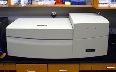
The Typhoon Trio+ instrument is a variable-mode imager that produces digital images of radioactive, fluorescent, or chemiluminescent samples. The Typhoon Trio+ instrument contains three internal lasers (blue, green, and red), with a scanning resolution of 10-μm pixel size.
- Versatile system platform, handles gel sandwiches, agarose and polyacrylamide gels, membranes, microplates and even microarrays
- Gels are scanned between glass plates, preventing drying and shrinkage and allowing further running and rescanning if required
- Powerful excitation sources and innovative high-quality confocal optics allow for the sensitive detection of low-abundance targets
- Red-, green- and blue-excitation wavelengths and a wide choice of emission filters enable imaging of an extensive variety of fluorophores
- Automated four-color fluorescence scanning allows multiplexing of multiple targets in the same sample ensuring accuracy of analysis, increasing throughput, and saving time
- Highly sensitive optics enable direct chemiluminescent imaging without intermediate exposure to films or screens
- Provides optimized detection of Cy2, Cy3, and Cy5 CyDye DIGE Fluor minimal and saturation dyes, enabling visualization of up to three differently labeled samples on a single gel
LI-COR Odyssey Fc Imager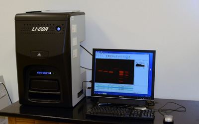
The Odyssey Fc System allows fast, easy image acquisition and analysis that eliminates film and the need for multiple exposures. It offers chemiluminescence detection and the benefits of two infrared fluorescence detection channels (700 nm, 800 nm) thus enabling quantitative two-color Western blot analysis. The Image Studio software allows accurate quantification over 6 logs of dynamic range.
Zeiss Axioplan 2 Microscope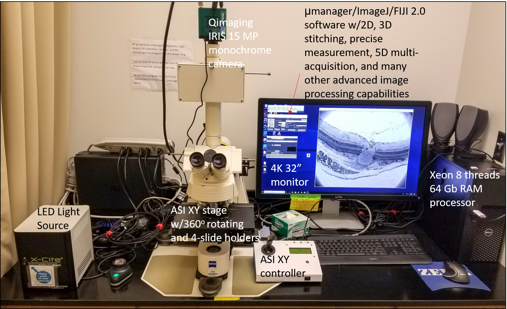
Combines brilliant optics and bright fluorescence, equipped with:
- Objectives (10X, 20X, 40X, 63X oil, 100X oil)
- Multichannel Fluorescence Filter (FITC, TRITC, Cy5, DAPI)
- Bright-field, Dark-field, Differential Interference Contrast (DIC)
- Zeis AxioCam HRm B/W and MRC-5 color cameras; photometrics IRIS9 B/W camera,
- ApoTome (pseudo-confocal) module
- Capable of time-lapse and Z-Stack acquisition mode and 3D deconvolution with axiovision software.
- Several versions of micromanager 64 bit open source software
- ImageJ and FIJI plugins
- ASI XY stage with controller
- Xcelitas X-Cite LEDboost light source
- 4k 32" monitor
Nikon AX-R Laser Confocal Microscope
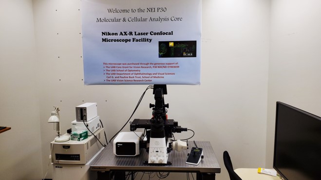
- 4 lasers for all commonly used fluors
- Large image stitching
- 2x to 100x objectives available including a 60x oil N.A. 1.49
- deconvolution and denoising in batch or individually
- Piezo Z-control
- color camera and led light source for color images
- x, y, z automated stage
- DIC and polarizing optics available
Return to Home Page of Ocular Phenotyping and Molecular Analysis Module