Molecular & Cellular Analysis Core
 |
 |
 |
| Core Director Steven Pittler, PhD pittler@uab.edu |
Associate Director Yuchen Wang, PhD wangyc@uab.edu |
Laboratory Manager José Luis Roig-Lopez, PhD joseluis@uab.edu |
Reserve your instrument time on your computer using the online scheduler http://scheduler.vsrc.uab.edu/molbio/ The Molecular and Cellular Analysis Core is housed in Volker Hall room 370. The core users are encouraged to turn off the lab lights when they leave especially after hours. And don't forget to log off the computers. All core users should acknowledge P30 EY003039 for funding. If you use the lab at all, please include this acknowledgment in every publication, poster and other scientific presentations. Please send a copy of any published works that cite the core and include in NCBI. That will help us to successfully compete for renewed funding. About The purpose of the Molecular & Cellular Analysis Core is to assist users with the molecular and cellular analysis of ocular tissues, the investigation of gene expression, including but not limited to primer and mutant design, amplification and quantitation of RNA or DNA products, cloning, and other molecular biology techniques. To accomplish that, the core acquired a comprehensive array of instrumentation. Day to day activities of the core are handled by laboratory director Dr. José Luis Roig-Lopez, who has extensive experience in many aspects of molecular biology, histology, and cell biology.
To facilitate the availability of these Core services, the Core has acquired comprehensive equipment
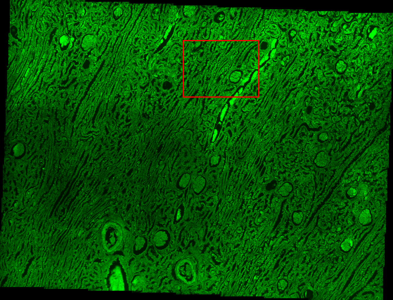
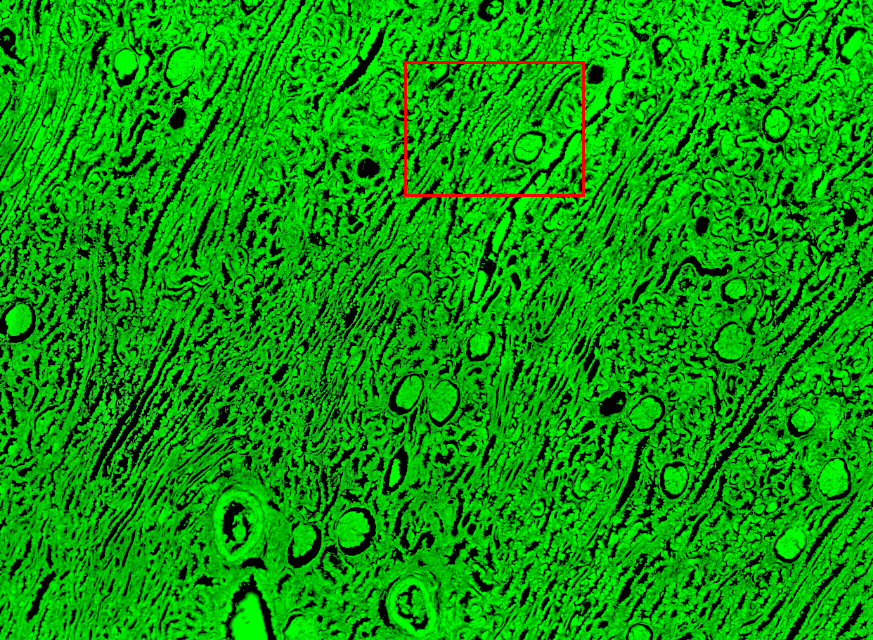
Fluo-1 test slide (bovine pulmonary artery endothelial cells)  The image was captured with µmanager V.2 software multi dimensional acquisition module controlling a Zeiss Axioplan 2 upright fluorescence microscope with a 20x objective, Excelitas LED120Boost light source, and a photometrics IRIS 9 MP CMOS camera with FITC, CY5, and DAPI filters. Exposure times were optimized manually. Useful links (some links may be available on campus only)
Primer design links:
|
||||||||