-
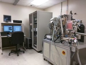
Computer Controlled Electrospinner
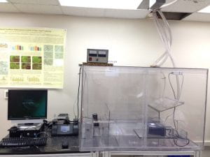
Computer Controlled Electrospinner
Automated electro spinners for making 3D-tissue-scaffolds from proteins, synthetic and biopolymers, blends
and composite nanofibers in tubular geometry of varying diameters ( 1 mm to 6 mm) and any lengths up to 30 cm for vascular grafts,
perfectly-aligned fiber scaffolds for nerve tissue engineering, bi-phasic and tri-phasic composite fibers (hydroxyapatite/ collagen/polymer
( PLA, PCL, PLGA, PLC)) fiberous scaffolds with 80-90 % porosity and interconnectedness with and without growth-factors for bone and craniofacial tissue engineering etc.
Reserve Equipment -
Diamond Microfabrication Lab
Diamond Microfabrication Lab
This facility contains two core systems.
A Sputter deposition system and a maskless lithography system provide researchers the capacity to create their desired patterns
in their choice of metals.AJA International Inc’s Polaris sputtering system has a 750 watts DC power source for the target gun
and a 100 Watts RF power source for creating RF plasma to clean samples. Metal films of thickness in the range 0.2 µm – 2 µm can be deposited using
the DC power source. Future upgrades planned for this system will allow for the deposition of both conducting and insulating thin films.
The maskless lithography system has the capability to create features with a resolution of 5 µm. The innovative smart filter on this system eliminates the
need for expensive physical masks for creating desired patterns on a given substrate. The microfabrication lab is equipped to carry out both wet-etch
and lift-off processes for creating patterns/circuits on planar and non-planar substrates.
Reserve Equipment -
Micro-Raman/Photoluminescence Spectroscopy
Micro-Raman/Photoluminescence Spectroscopy
Raman spectroscopy is a powerful technique for probing the local atomic environment in a wide variety of materials
by detecting the phonon vibrations through the material. Particularly sensitive for carbon-based materials.
It is complementary to the FT-IR technique.
Reserve Equipment -
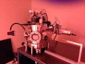
Microwave Plasma Chemical Vapor Deposition
Microwave Plasma Chemical Vapor Deposition
The 1.2 KW, 6 KW Microwave Plasma Chemical Vapor Deposition systems are utilized for growing single crystal and nanocrystalline diamond
on various substrates such as diamond, silicon, and titanium alloys. Shown to the left is the 1.2 KW CVD system.
This system is primarily used for growing homoepitaxial diamond for the purpose of fabricating designer diamond anvils
utilized in high-pressure research.
Reserve Equipment -
Multi-Axis Wear Simulation Test Apparatus
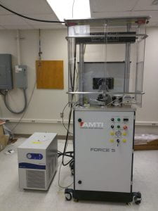
Multi-Axis Wear Simulation Test Apparatus
The Force-5 (AMTI, Inc.) wear simulation instrument for the testing of materials for biomechanical applications, such as the evaluation
of joint replacement implants. The machine can be set up to test various types of devices such as hips, knees, and temporomandibular joints.
The motion can be programmed to mimic the complex motion of these implants and uses feedback to control the motion.
With a maximum vertical load of 1000lbs. and 4 independently controlled axes of motion, the machine can replicate patterns of motion such as walking
or chewing using a user-configurable computer control. A six-axis load cell gathers force and displacement data for analysis.
The joint in testing can be maintained in a temperature-controlled, fluid-filled environment for proper biomechanical considerations.
Reserve Equipment -
Panalytical Empyrean Multi-purpose X-ray diffractometer
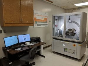
Panalytical Empyrean Multi-purpose X-ray diffractometer
The Panalytical Empyrean Multi-purpose X-ray diffractometer has the capability to analyze many types of samples, including powders,
thin films, and epitaxial films. Samples can be analyzed in reflection or transmission and the instrument is outfitted with multiple stages
including a 3-axis stage and a reflection/transmission spinning stage. Other types of analysis include small angle X-ray scattering (SAXS),
micro-diffraction (~300um spot size), and high-resolution single crystal analysis, such as pole figures and reciprocal space mapping (RSM).
The instrument has two detectors: an array detector for fast powder diffraction scans and RSM as well as a standard proportional detector
with a parallel plate collimator for thin film scans. Multiple optics are available including the BBHD (standard optic with beta filtering),
a monochromator for high resolution scans, a focusing mirror for SAXS and transmission scans, and the micro-diffraction optic.
Reserve Equipment -
Scanning Electron Microscope (SEM)
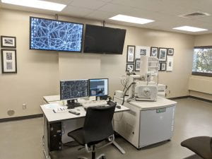
Scanning Electron Microscope (SEM)
The Quanta 650 FEG scanning electron microscope (SEM) was purchased in 2012 with an NSF-MRI grant for $550,000 and is located
at the School of Engineering. The SEM provides high resolution images with resolution down to the nanometer range.
It can image samples ranging from metals to biological samples and even wet materials. It also can perform energy-dispersive x-ray analysis (EDAX)
to provide elemental composition. Non-conducting samples can be imaged with the aid of the new sputter coater which deposits a
thin layer of gold-palladium on the surface to improve imaging.
Reserve Equipment -
Spectrofluorometer with microscope coupling optics
Spectrofluorometer with microscope coupling optics
The Horiba Scientific Fluorolog 321 double grating spectrometer with TE-cooled photomultiplier will be used for high sensitivity
fluorescence detection upon Troponin adsorption. This system has a solid sample and custom microscope coupling optics/slits
to allow fluorescence mapping at the sub-micron level using an attached Zeiss fluorescent microscope with custom camera
and spectrometer interface components.
Reserve Equipment -
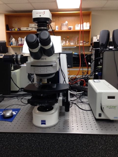
Fluorescent Microscope
Fluorescent Microscope
The Zeiss Axio Imager A2 fluorescent microscope has both reflective/transmission imaging capability and is fiber-optically coupled to the Horiba spectrophotometer for solid sample fluorescence measurements from sub-micron areas.
Viscometer: The Brookfield Engineering LVDV-II+PRO Cone/Plate viscometer system allows rheological measurements of small sample volumes as low as 0.5 ml. This will enable us to study mixing and viscosity of polymer blends and composites needed to print “inks” for DPN.
Plasma and UV/ozone cleaners: The Harrick plasma cleaner and Novascan 4” UV/ozone cleaner are used to remove surface contamination necessary for controlled DPN experiments and also provides manipulation of surface wettability.
Reserve Equipment -
X-ray Photoelectron Spectroscopy Lab
X-ray Photoelectron Spectroscopy Lab
The core technology of the VersaProbe 5000 is PHI’s patented, monochromatic, micro-focused, scanning x-ray source
which provides excellent large area and superior micro-area spectroscopy performance. Spectroscopy, depth profiling,
and imaging can all be performed over the full range of x-ray beam sizes including the minimum x-ray beam size of 10 μm.
Multi-technique options include chemical mapping, quantitative elemental analysis, a hot/cold sample stage, and sample treatment chambers.
Reserve Equipment