Mission Statement
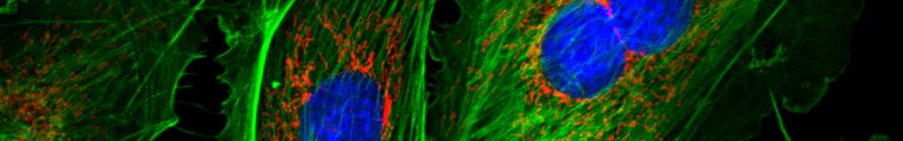
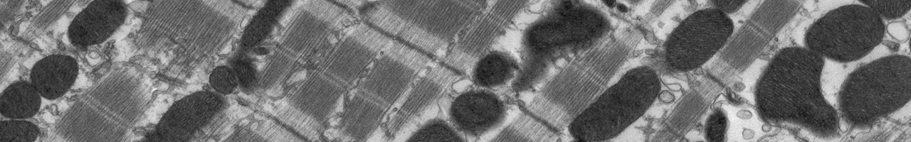
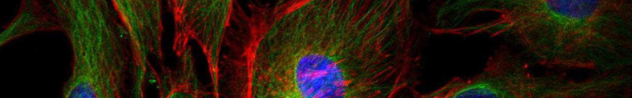
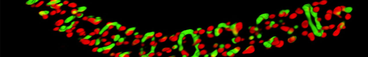
The High Resolution Imaging Facility (HRIF) provides state-of-the-art imaging resources and technical support to the UAB community. HRIF offers electron microscopy and light microscopy including confocal, live cell, multi-photon, widefield, super resolution, and image analysis. To effectively implement these technologies, we provide consultations, expert training, and support for all our systems. We are open to all UAB investigators as well as other investigators in the research community.


HRIF News & Events
-
UAB's Core Day 2026

-
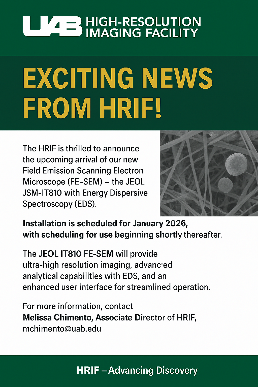
Arriving Soon: Field Emission Scanning Electron Microscope (FE-SEM)
-
4 cutting-edge machines powering UAB discoveries
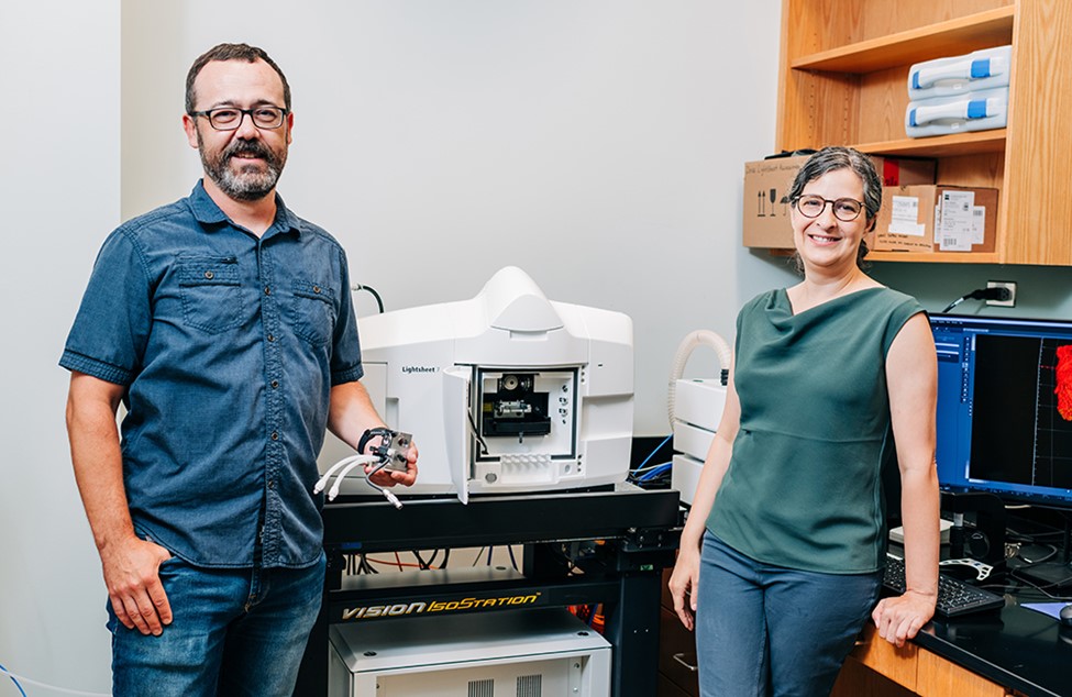
Photo by ANDREA MABRY | University RelationsAlexa Mattheyses, Ph.D. (right), director of the UAB High-Resolution Imaging Facility, and Robert Grabski, Ph.D., research associate in the High-Resolution Imaging Facility. They are pictured with the Zeiss Lightsheet 7 light sheet microscope, an advanced imaging device capable of fluorescent imaging of living cells and tissues.
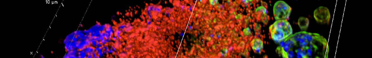
What it does: A light sheet fluorescent microscope, or LSFM, allows researchers to visualize the relationships between cells and proteins in intact tissues, organs and organisms at high volume and at a range of sizes and resolutions. Because of its image acquisition speed, the Zeiss Lightsheet 7 can perform data collection from large samples rapidly and efficiently, “allowing us to image far larger samples than we are currently capable of, including whole organisms and intact cleared tissues,” Mattheyses wrote in her grant abstract. Light sheet fluorescent microscopy also “substantially reduces photobleaching and photodamage,” she added. Samples do not need to be mounted on a slide or coverslip, “allowing more physiological imaging of model organisms and intact tissues,” Mattheyses said. “Finally, samples can be rotated relative to the imaging plane, allowing ideal orientation and collection of multiple views.”