
Lightsheet Videos
Mouse Embryo with fluorescence in green and red. Video was generated using Imaris 10.2 (Oxford Instruments). Imaging was performed using Zeiss Lightsheet 7 at High Resolution Imaging Facility, UAB, by Dr. Robert Grabski. Original research and sample preparation by Dr. Jianbo Wang, Department of Cell, Developmental, and Integrative Biology, UAB.
The video was generated using Imaris 10.2 software (Oxford Instruments) from a cleared mouse brain tissue. Imaging was performed using Zeiss Lightsheet7 microscope by Dr. Robert Grabski at High Resolution Imaging Facility, University of Alabama at Birmingham. Original research and sample preparation by Dr. Naoki Hama and Dr. Masakazu Kamata, Department of Microbiology, University of Alabama at Birmingham.
TEM Images (FEI Tecnai T12)
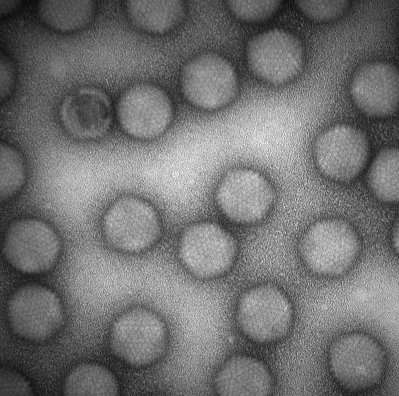
Adenovirus
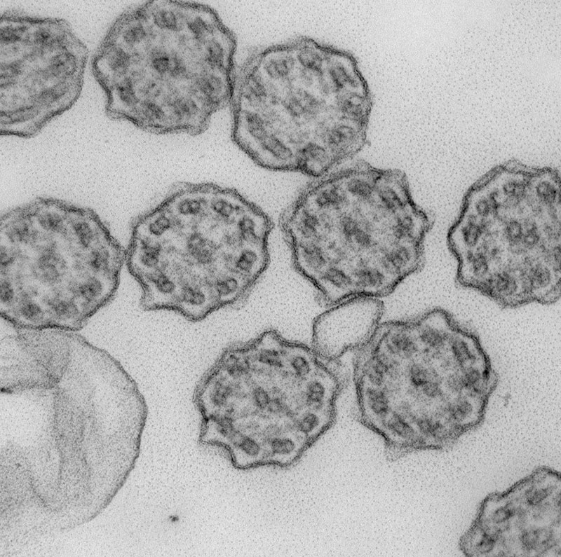
Cilia
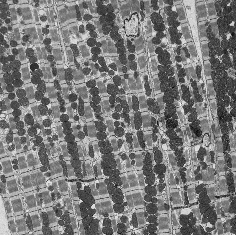
Mouse heart
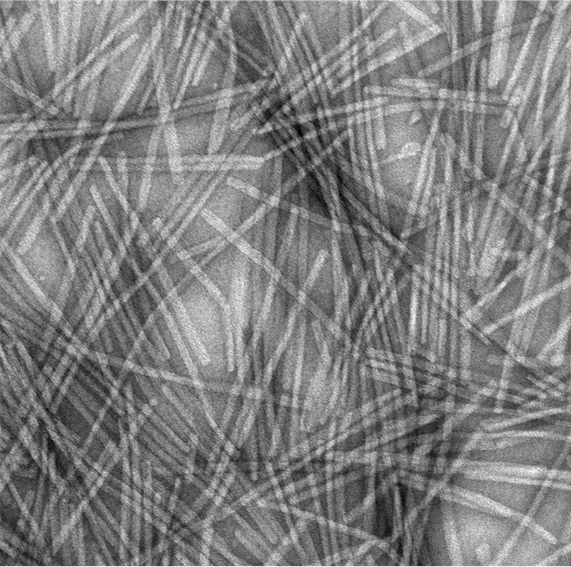
Carbon nanofibers
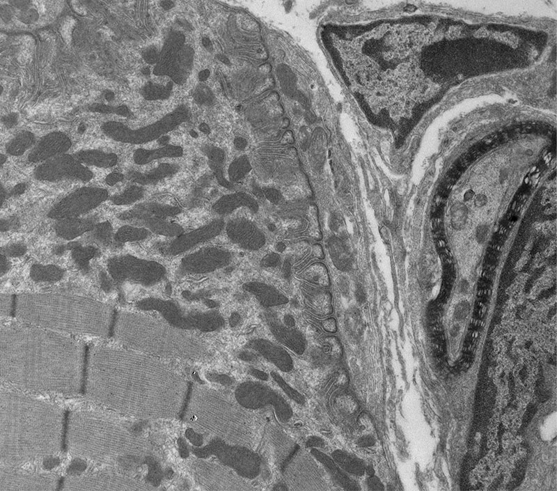
Neuromuscular junction
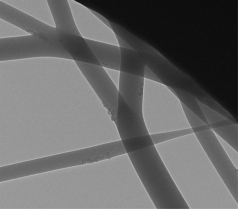
Electrospun fibers
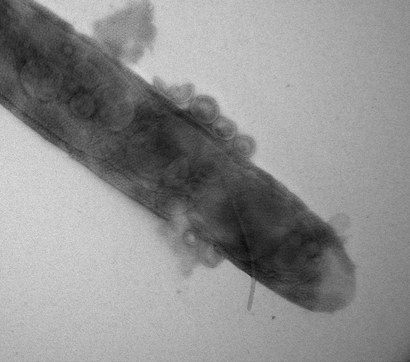
Local pond water
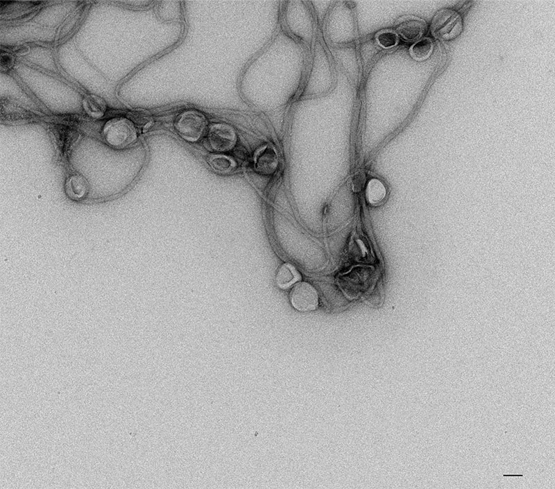
Collagen and exosomes
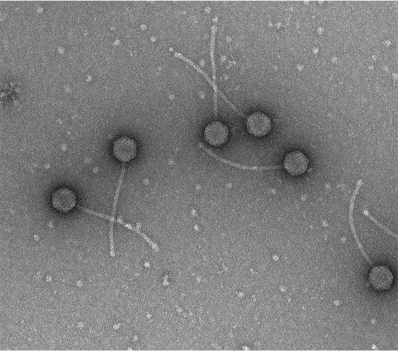
Bacteriophages
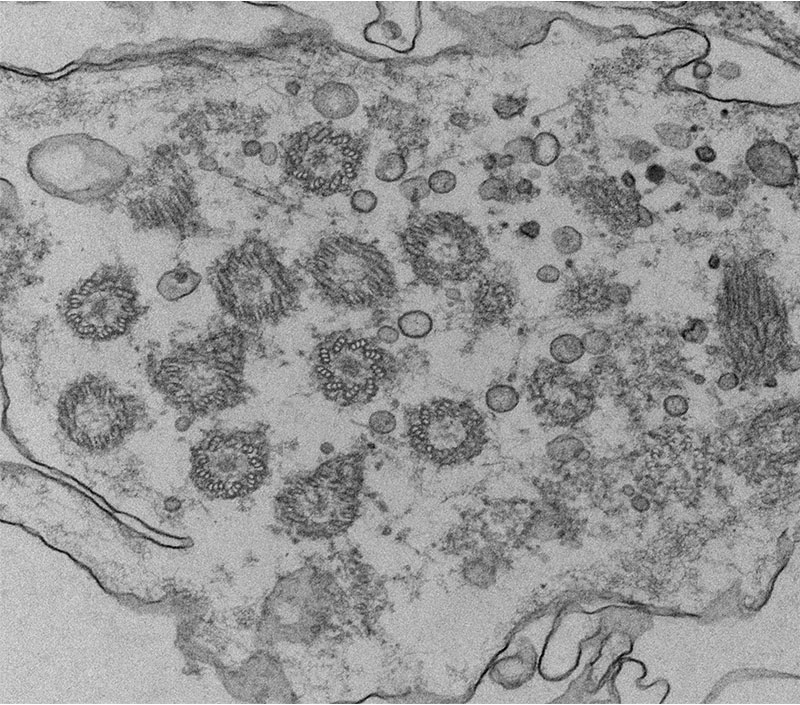
Cilia
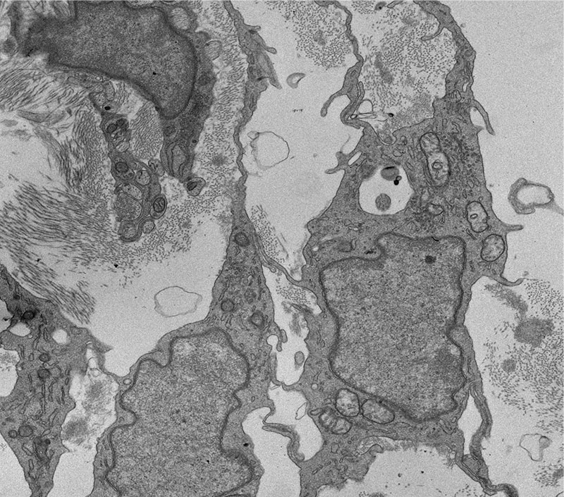
Ciliary muscle
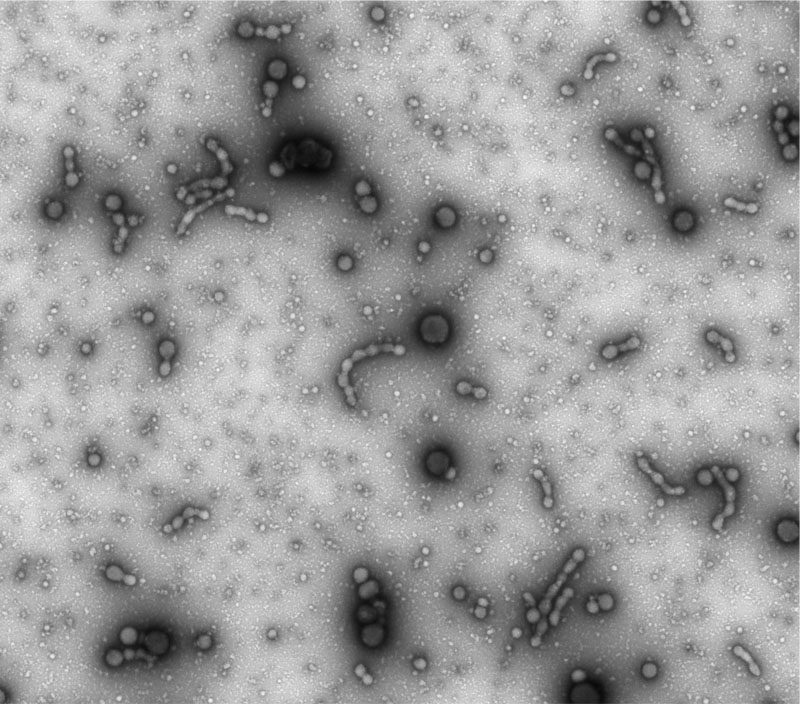
Micro vesicles
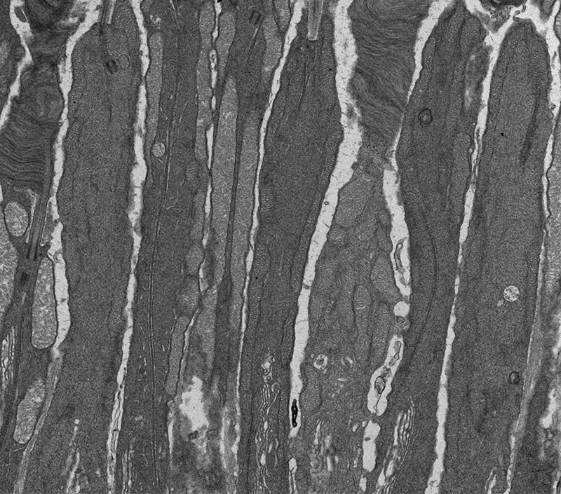
Mouse photoreceptors
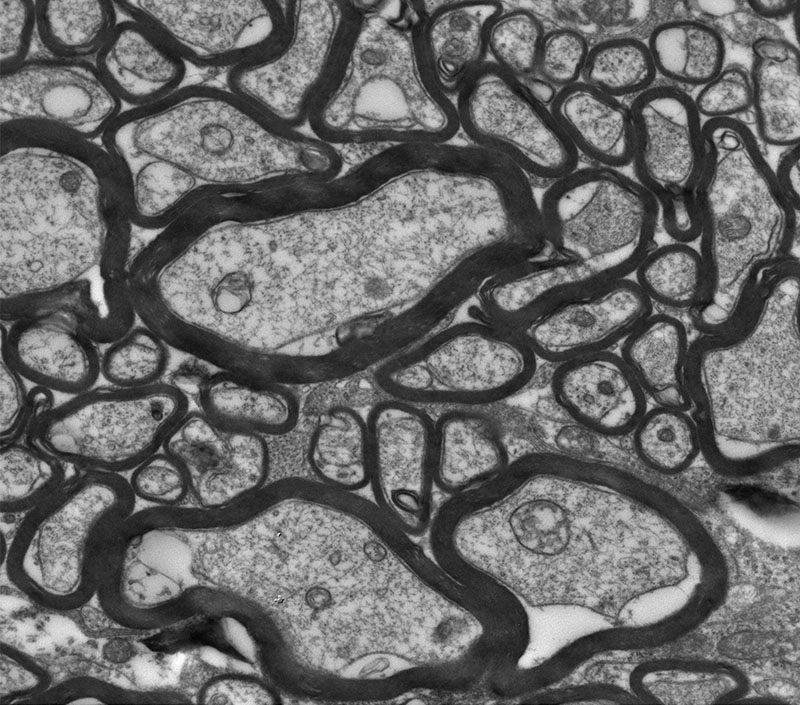
Myelin
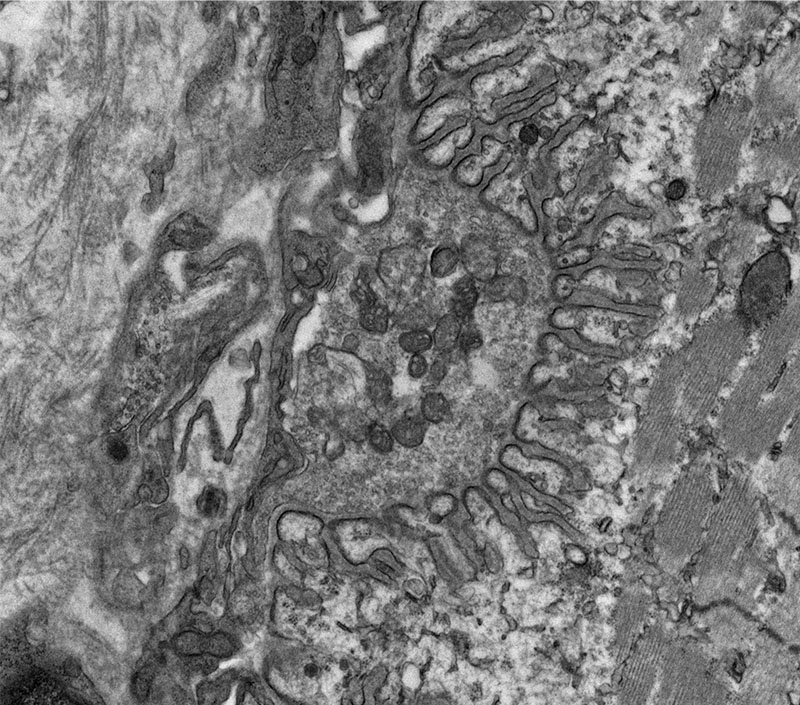
Neuromuscular junction
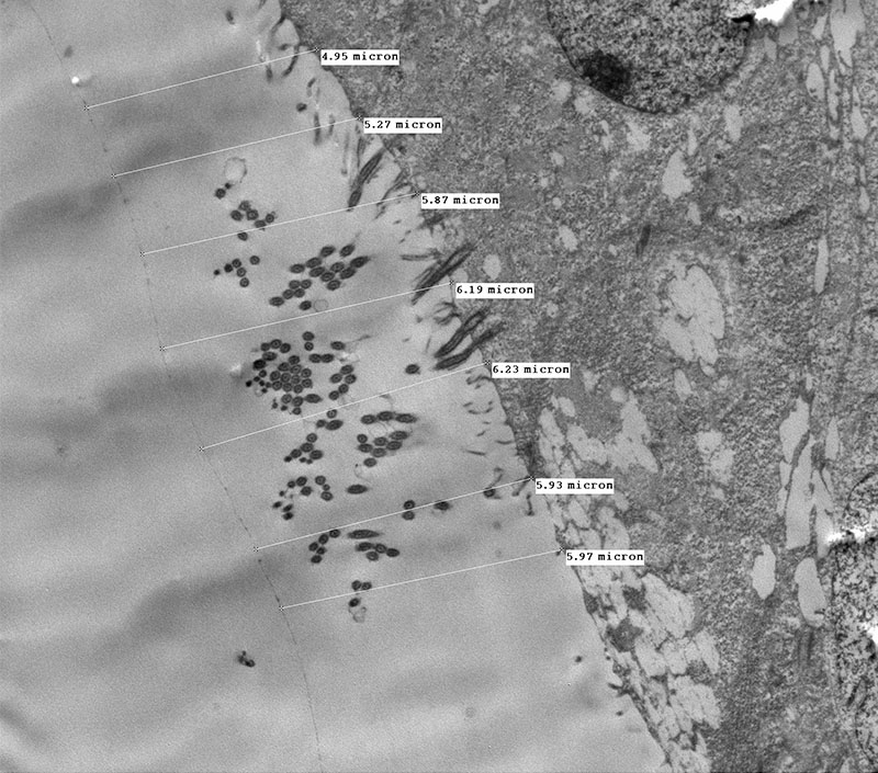
Lung periciliary layer
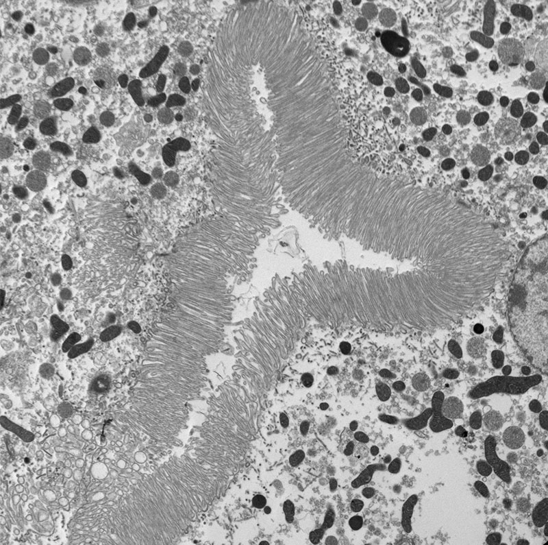
Mouse kidney
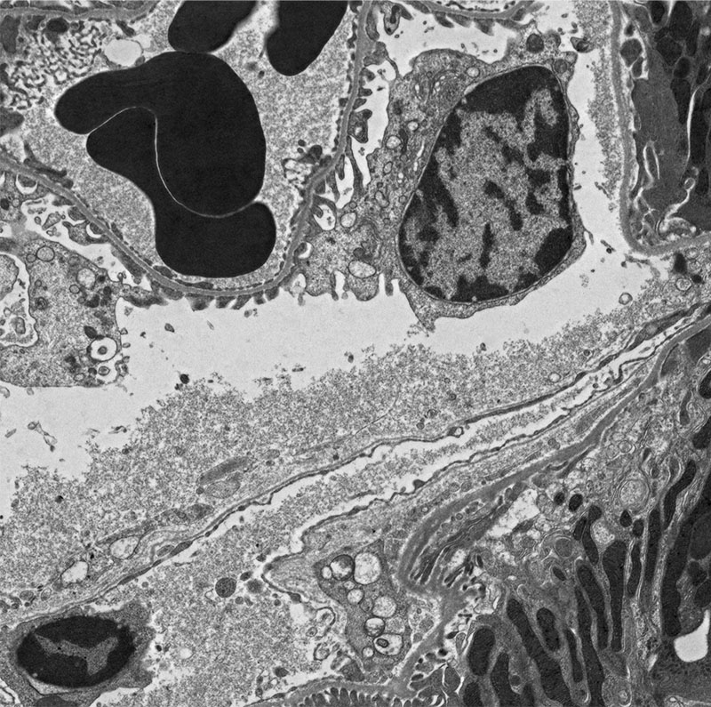
Mouse kidney
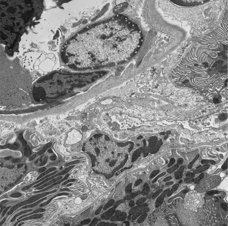
Mouse kidney
Fluorescence Images (Various)
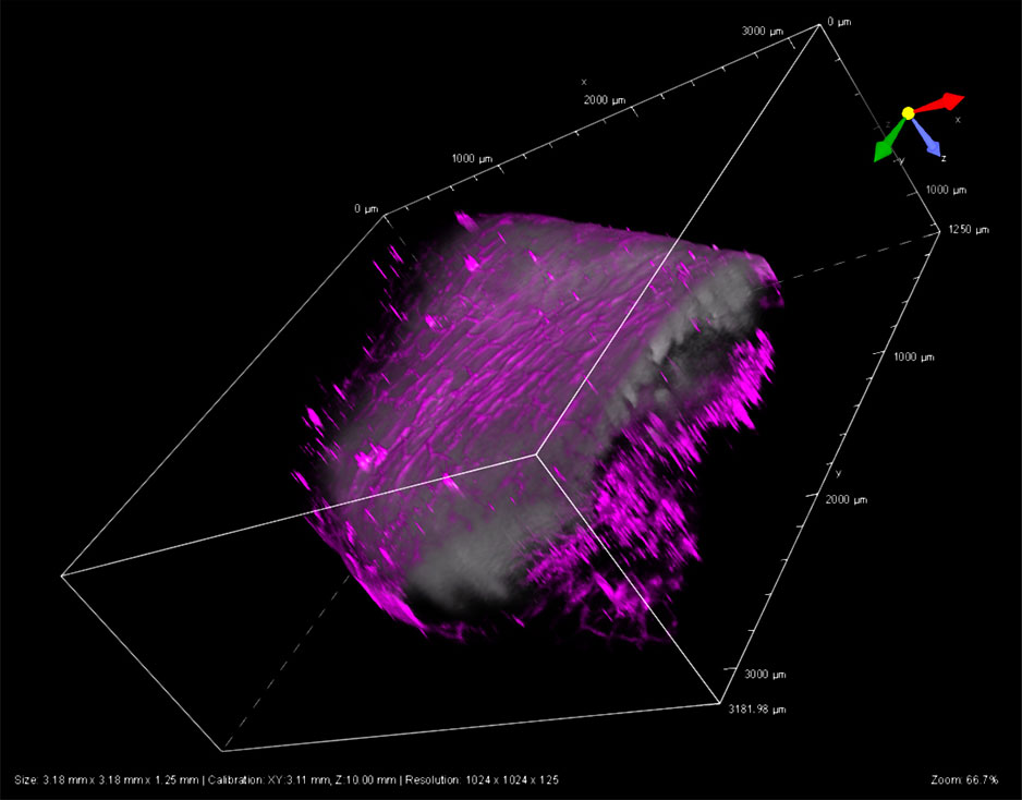
Mouse Intestine
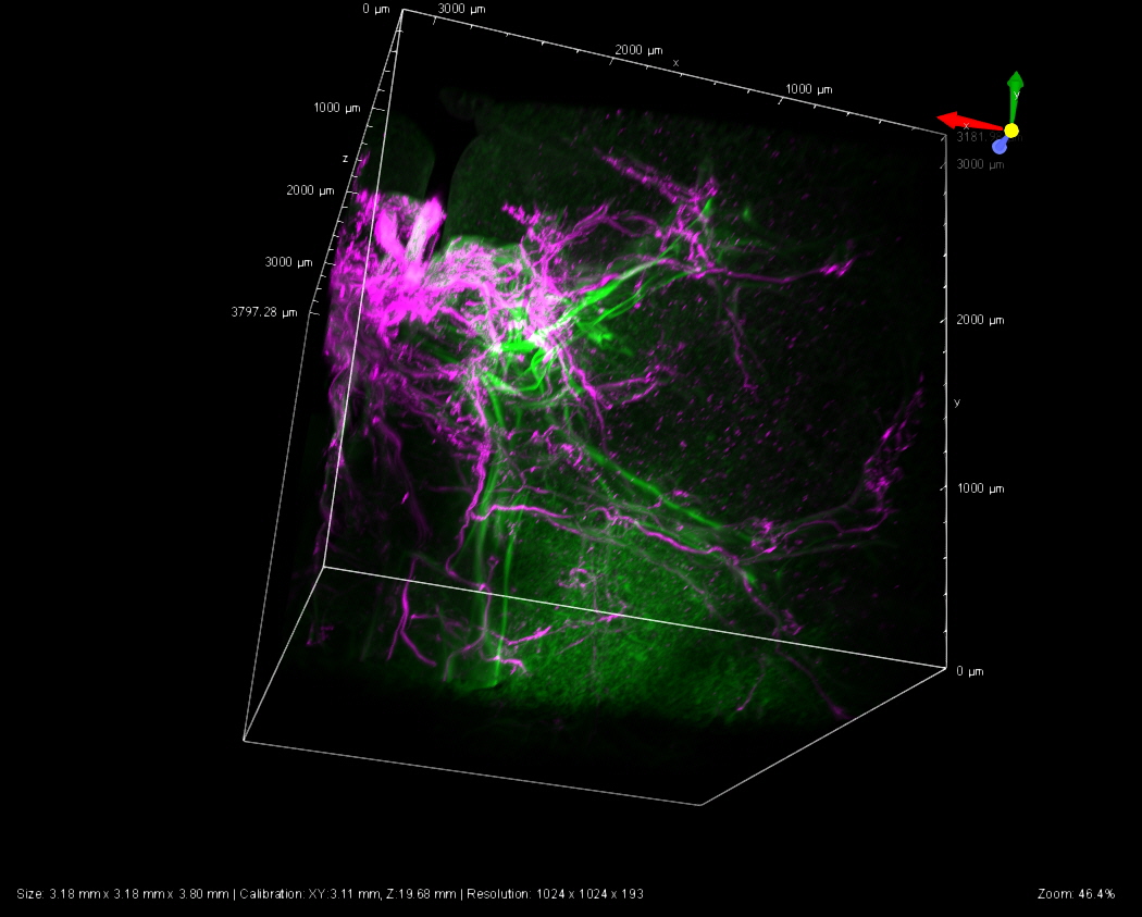
Mouse Lung 3D
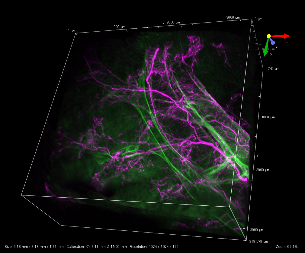
Mouse Lung 3D
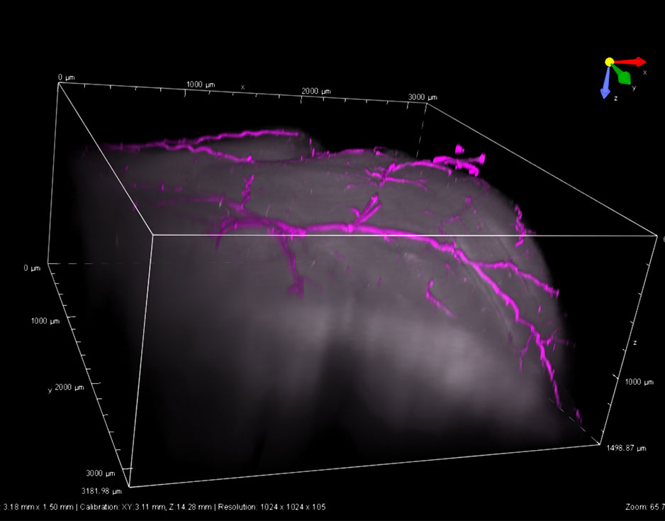
Mouse Brian
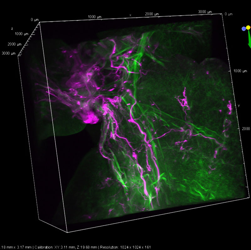
Mouse Lung 3D
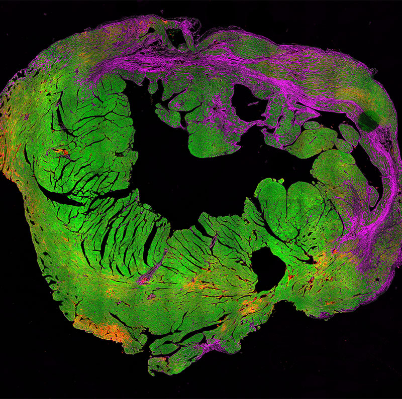
Mouse Heart
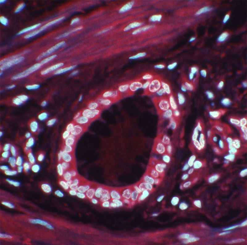
Liver
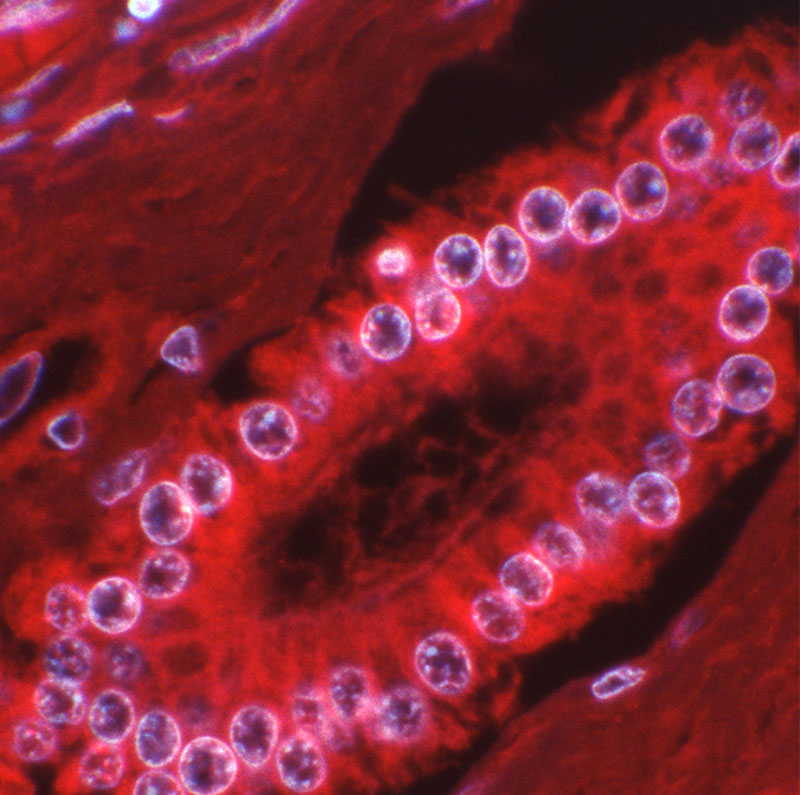
Liver
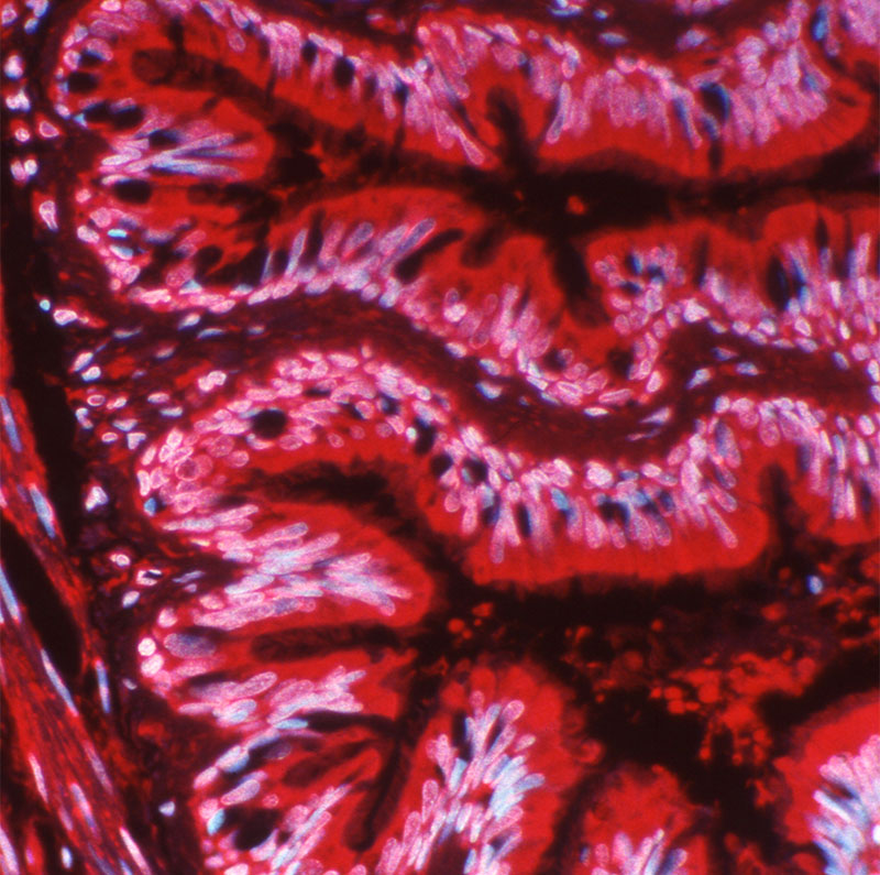
Lung
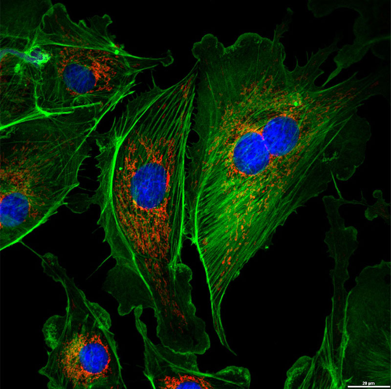
BPAE cells
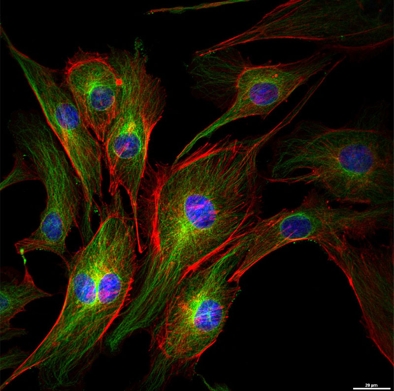
BPAE red actin
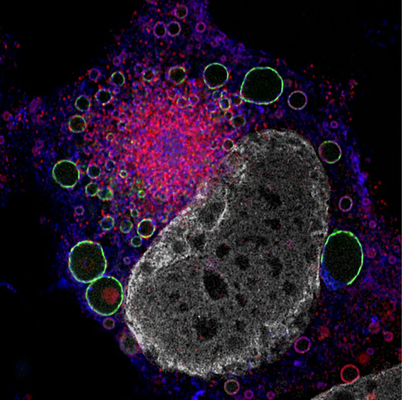
Fibroblast infected w/ HCMV
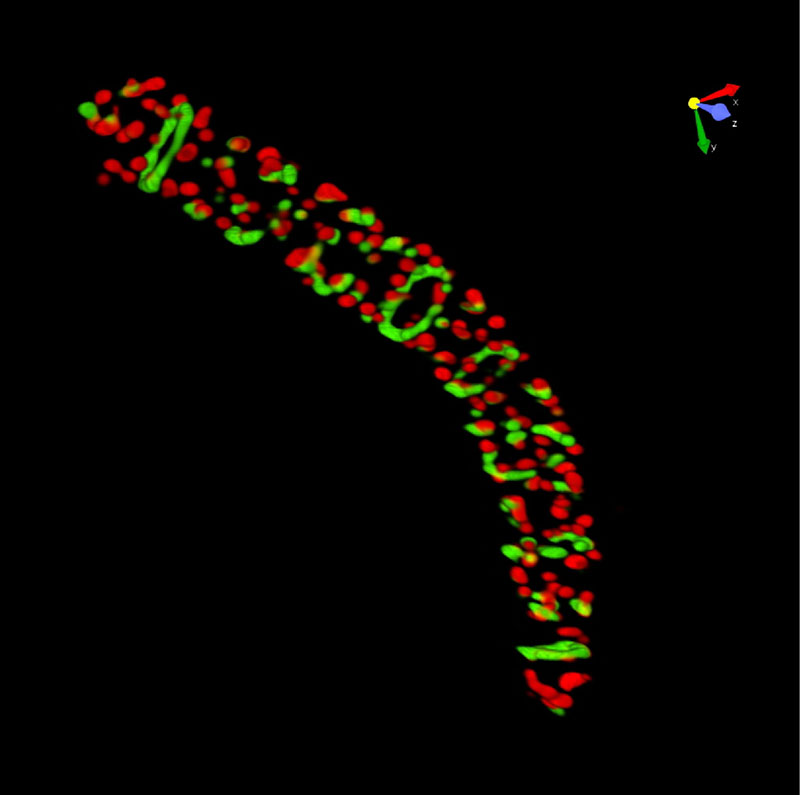
Bacteria
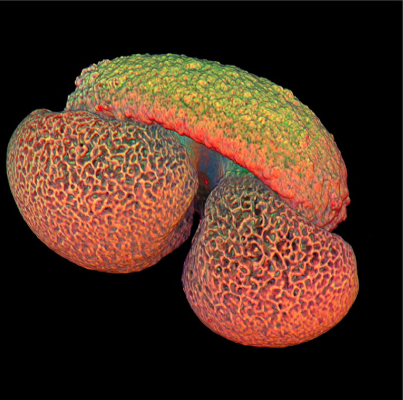
Pollen
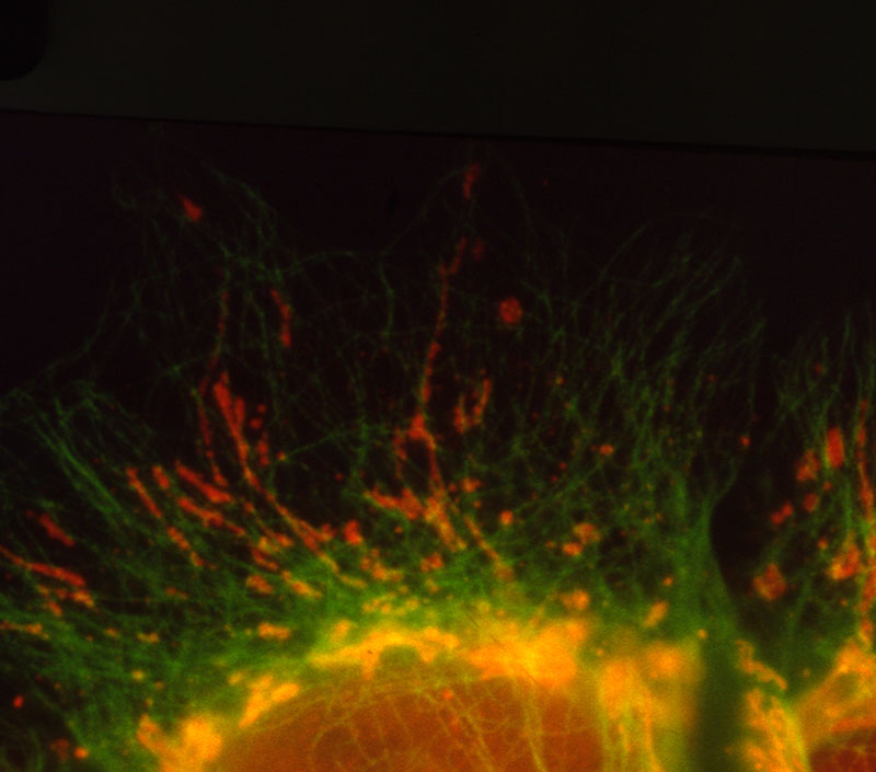
Cytoskeleton and mitochondria
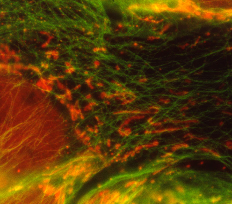
Mitochondria
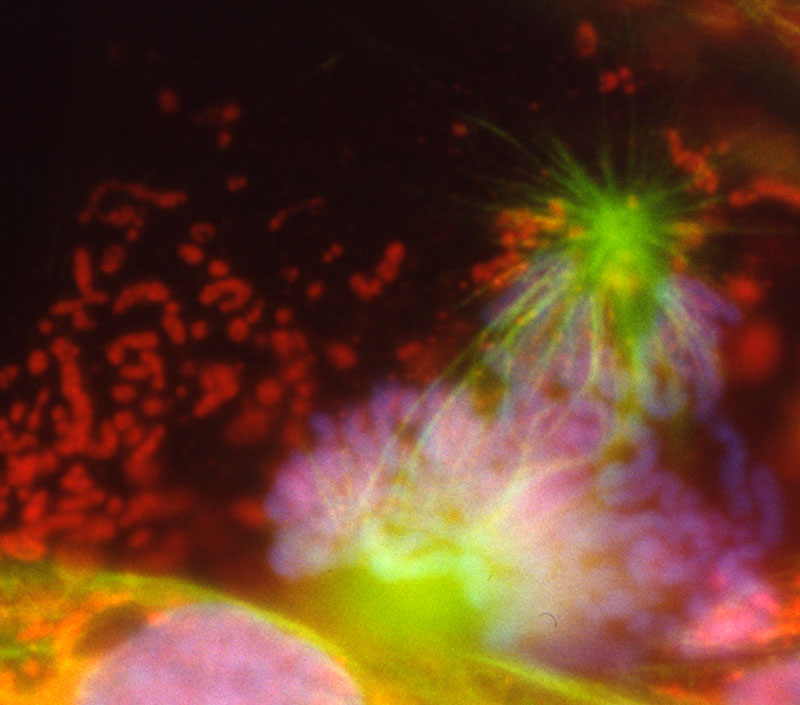
Mitotic cells
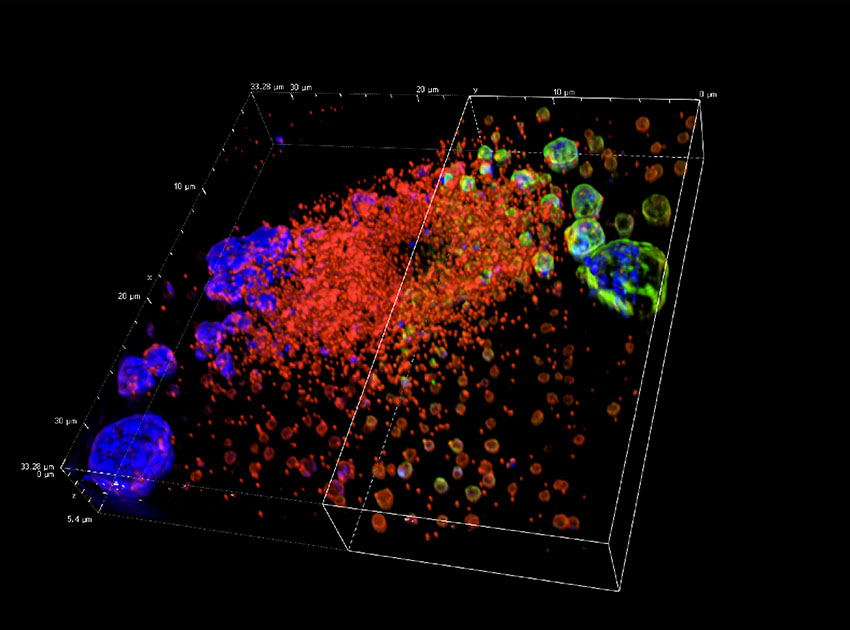
SIM HCMV
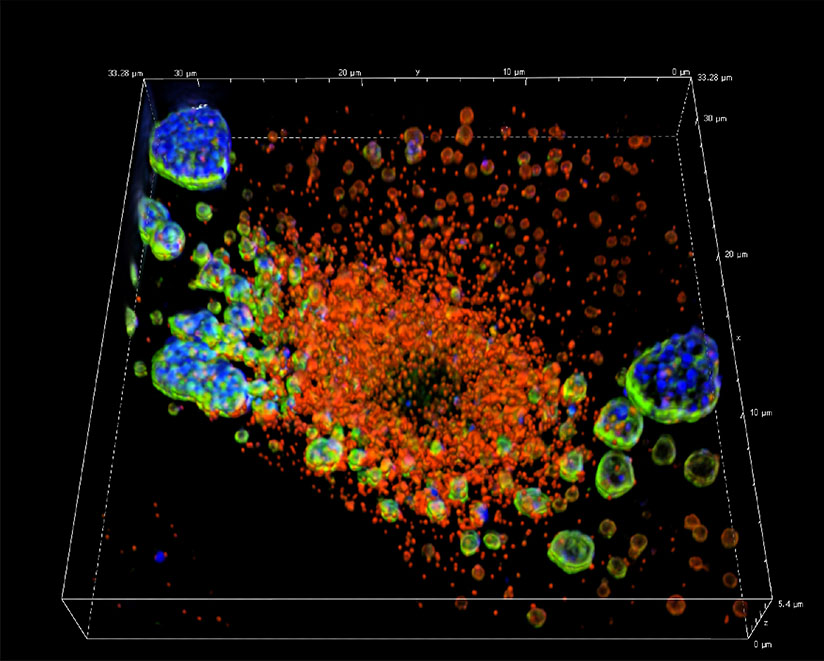
SIM HCMV 2
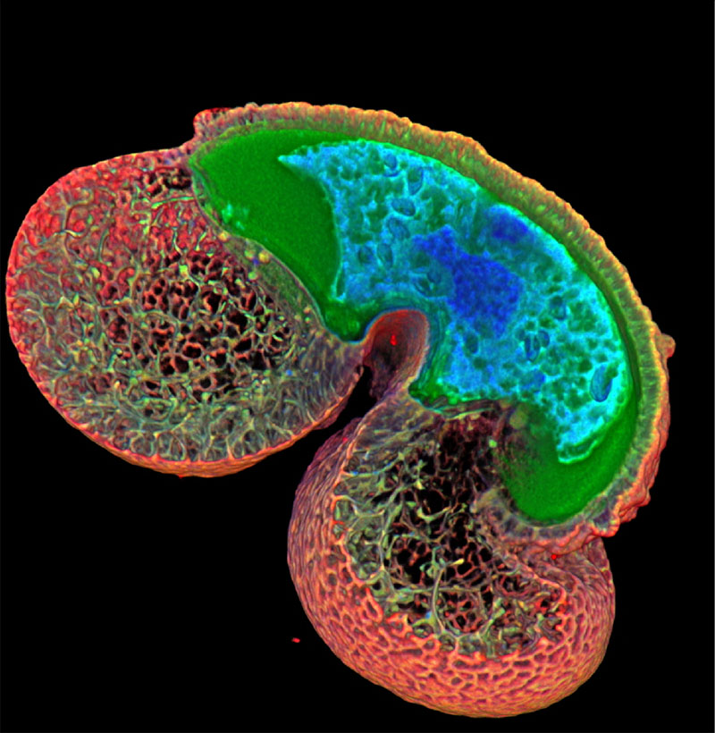
Polen2
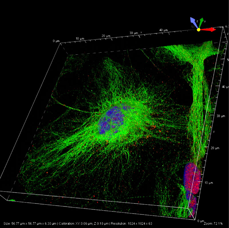
Nestin