 |
Gorgas Case 2002-08 |
 |
|
The following patient was seen by the 2002 Gorgas Class in the outpatient clinic of the Tropical Medicine Institute.
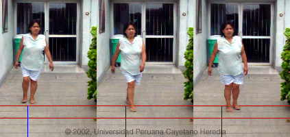  History: 39 year old woman with 8 years of difficulty walking, parasthesias in both legs, and a feeling that her legs were hard and weak. She stumbles frequently and trips on her own feet. For the past 3 years she has had lumbar pain, increasing episodes of loss of bladder control and frequent constipation. Recent onset of foreign body feeling in both eyes but no loss of vision. Epidemiology: Born and living in Lima. Healthy parents, normal delivery, was breastfed. Five healthy children. Monogamous with husband of African-Latino American origin who died accidentally 2 years before the diagnosis was made. No surgery, blood transfusions, IV drug use or STDs. 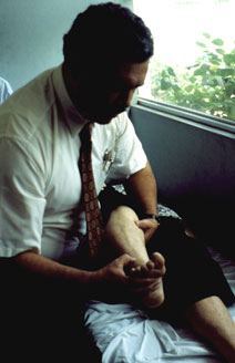 Physical Examination: Afebrile. No organomegaly or lymphadenopathy. Bilateral spastic paraparesis with symmetrical hyper-reflexia of legs and bilateral ankle clonus (being demonstrated by Dr. Gotuzzo in image at right). Upgoing toe on right. Slow spastic scissors-like gait characteristic of this disease is demonstrated in the images above. The patient can walk only by swinging one leg around and directly in front of the other. The back leg is then brought alongside. This results in forward movement with a sideways component to each step, that alternates sides with each successive step. Labs/X-ray: Hb 13.5, WBC 7,200 normal differential. Renal, hepatic function normal. FTA-abs-negative. PPD 5 mm Chest x-ray normal. Spinal plain films normal. |
|
Diagnosis: Tropical spastic paraparesis (TSP) secondary to HTLV-1 infection. Also called HTLV-1 Associated Myelopathy (HAM).
Discussion: ELISA and western blot were positive for HTLV-1. Her mother was HTLV-1 negative and her father and husband (died 2 years before the diagnosis) were not tested. Our previously published case series (J Neurol Sci 1996;143:114-7) indicates breastfeeding to be the major mode of transmission in Perú, but in this case it appears to have been acquired sexually, which is an also well-described risk along with blood transfusion and IV drug abuse. Other conditions associated with HTLV-1 infection are acute T-cell leukemia/lymphoma (normal blood film in this patient), crusted (Norwegian) scabies, strongyloides hyperinfection, and autoimmune disease including Sjogrens, and thyroiditis. The patient's 15 year old daughter is HTLV-1 positive and has chronic infectious dermatitis (Quervain's dermatitis). The prevalence of HTLV-1 in South America is generally under-appreciated, normally being associated with Japanese, Caribbean, and African populations. In Perú, the disease is highly endemic (2-3% seropositivity) in Andean areas of the country, in Quechua populations who have had no contact with Japanese immigrants to the country. This patient likely acquired her infection from her husband. High prevalence rates have also been described in the relatively small African-Latino American population in Perú. Other South American countries with significant rates of HTLV-1 include Brazil (0.45% of all blood donors), Colombia, and Ecuador. The clinical syndrome of TSP had been found fairly commonly in the Caribbean, Africa, and South America for over a century. Clinical features characteristically indicate a myelopathy at the level of the thoracic cord. The syndrome includes progressive bilateral spastic paraparesis, hyper-reflexia (often includes clonus and Babinski's sign), and sphincter signs manifest most often by urinary bladder dysfunction. Upper extremity spasticity is uncommon but occasionally there may be a Hoffman's sign. Parasthesias are common but objective sensory findings are present in only about 20% and manifest usually by altered vibration sense up to a thoracic lesion level. Cranial nerve and cognitive deficits are not present. Until 1985 TSP was thought to be mostly idiopathic or attributable to malnutrition, syphilis, metabolic disorders/toxins, or extrinsic cord compression. Multiple sclerosis is a disease of temperate, not tropical, climates so is usually easy to exclude in tropical countries. MS is often accompanied by optic neuritis and usually waxes and wanes at the outset in contrast to TSP, which is chronically progressive. Beginning in 1985 it was found in various HTLV-1 endemic areas that from 60-100% of TSP cases were associated with HTLV-1 infection. A similar clinical syndrome in Japan was also found to be due to HTLV-1 in a temperate climate and named HAM. Although this is clearly the same clinical process, for unclear reasons the lifetime risk of TSP/HAM in HTLV-1 infected individuals is about 2-4% in Latin America/Caribbean and about 0.025% in Japan. Age of onset is usually in the 3rd decade, the female predominance of TSP is about 2:1, and 20-30% are wheelchair bound within 10 years of onset. CSF usually shows increased protein, small numbers of mononuclear cells, and demonstrable HTLV-1 antibodies. MRI, when available, will usually show lesions of perivascular demylelination in the thoracic cord. Extrapolation of prevalence data in blood donors and the 2-4% lifetime risk of TSP found in the smaller studies indicates that there may be tens of thousands of cases of TSP in Latin America alone. HTLV-1 is CD4 tropic. Studies indicate TSP patients have higher pro-viral loads than non-TSP HTLV-1 patients and have high levels of cytotoxic anti-HTLV-1 lymphocytes. It is hypothesized that cytotoxic T-lympocytes attacking HTLV-1 infected CD4 cells in the spinal cord in patients with high viral loads may be responsible for the demyelinating lesions of TSP. Corticosteroid therapy is generally ineffective in these patients and some preliminary trials with anti-retroviral agents are underway. |