 |
Gorgas Case 2012-02 |
 |
|
The following patient was seen in the outpatient department of the 36-bed Tropical Disease Unit at Cayetano Heredia National Hospital.
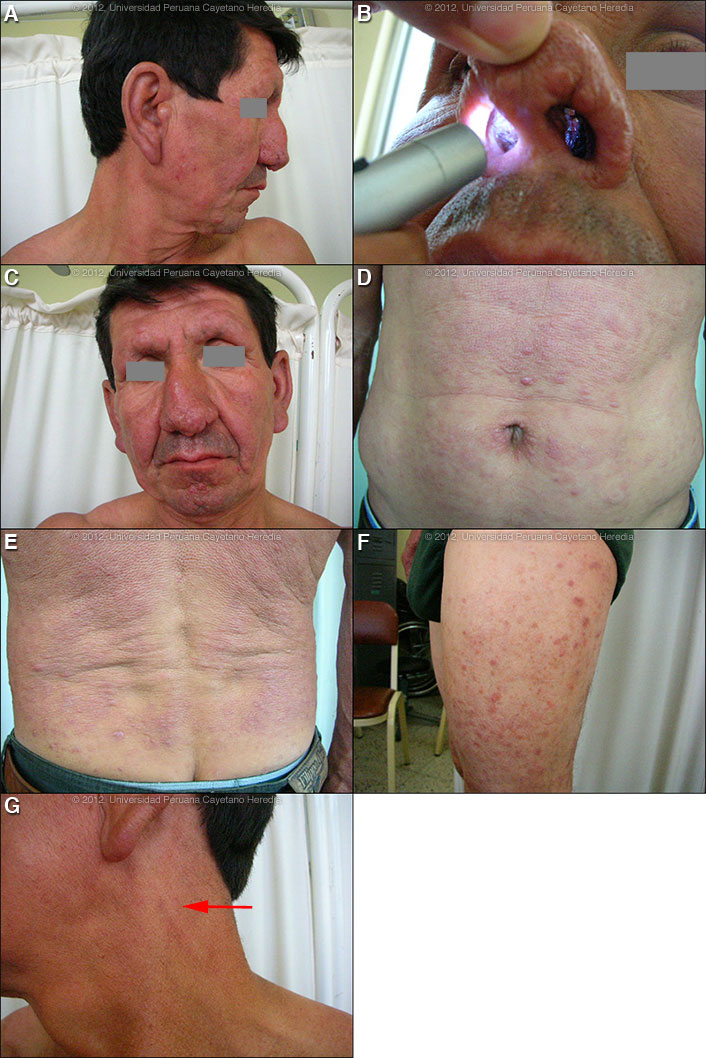
 History: 47 year old man with a 10 month history of self-described “hives” which appeared first on his abdomen, then the waist, lumbo-sacral region, the knees and later on his upper limbs and face. The lesions are non-pruritic and painless. He also had nasal congestion and nose bleeds. He denies other symptoms. He received various anti-allergic therapies without improvement. No relevant past medical history. Epidemiology: Farmer from rural Amazonas Department. Unmarried and lives alone. Physical Examination: Afebrile. HEENT: thickened earlobes and alae nasae with inflamed, infiltrated, eroded, nasal cartilage with nasal septal perforation [Images A, B]. Pharynx clear, normal oral mucosa. Chest clear, no hepatosplenomegaly or lymphadenopathy. Skin: diffuse, symmetric, erythematous cutaneous infiltrate on the face, except the axillae, midline of the back scalp, palms and soles. Multiple non-tender papules on the lips, chin, thorax, waist and limbs, and some small superficial nodules [Images C, D, E, F]. Normal scalp hair but loss of eyebrows (madarosis) and some eyebrow loss. Neurologic Exam: Bilateral enlargement of great auricular [Image G, arrow], ulnar, common peroneal and posterior tibial nerves, with impairment of sensation on hands, forearms, legs, feet as well as over the skin lesions. In addition, mild loss of motor function in the distribution of both ulnar nerves, right median nerve and right common peroneal nerve. Laboratory Results: Hematocrit: 42%, WBC: 6000 (55% neutrophils, 1% eosinophils, 1% basophils, 10% monocytes and 33% lymphocytes), Platelets: 319000, Glucose: 92mg/dL, Albumin: 3.9 g/dL, AST: 31 U (normal), ALT: 15 U (normal). Normal urine.
|
|
Diagnosis: Mycobacterium leprae. Multibacillary leprosy according to the WHO classification. Lepromatous leprosy (LL) leprosy according to the Ridley-Jopling classification.
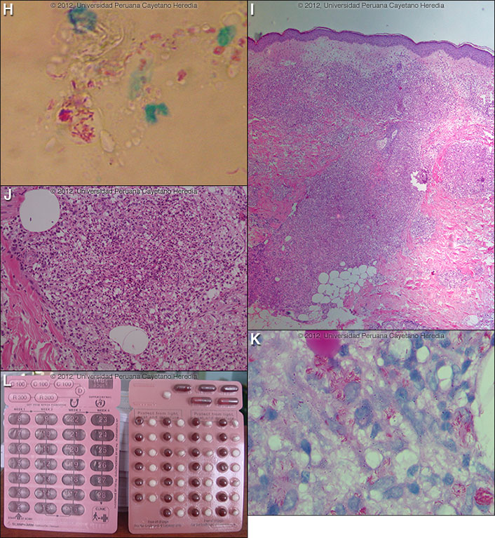
Discussion: Slit skin smears from earlobes, elbows (laterally to avoid the olecranon) and knees (laterally) showed 6+ bacilli/per high-powered field with globi [Image H]. Slit skin smears are performed by making small (5 mm length, 2 mm depth) slits in pinched skin (to avoid bleeding), the edges of which are scraped. The material obtained is smeared on a clean slide and stained for AFB. Generally ear lobes, elbows, and knees are examined. The bacterial index ranges from zero (no bacilli in 100 oil-immersion fields) to 6+ (many clumps or globi or over 1000 bacilli in one field). Skin biopsy [Images I, J]. The low power view shows attenuated epidermis, a clear sub-epidermal zone with edema of the papillary dermis and a widespread inflammatory infiltrate throughout the dermis. Deep in the dermis the infiltrate follows blood vessels and nerve fibers. The high power view shows that the infiltrate consists mainly of mononuclear cells (macrophages) many of which are vacuolated; there are also areas of infiltration with neutrophils. Under oil-immersion, the Fite Faraco Acid-Fast stain of tissue [Image K] shows abundant intracellular bacilli with many in clumps and globi. The changes are consistent with lepromatous leprosy in reaction. The linear distribution of the infiltrate (following nerves and blood vessels), and the foamy characteristic of the histiocytes are an important clue to the diagnosis of leprosy. The presence of neutrophils and the edema of the papillary dermis are also an important clue to a Type 2 reaction (see discussion below). Type 1 reactions, by contrast, are lymphocyte rich. Leprosy is a disease of peripheral nerves and skin. Leprosy can be diagnosed clinically in any patient with simultaneous skin lesions and sensory loss over the lesions unless there is hyperkeratosis. However, in early lepromatous cases, sensation is normal over the lesions. Thus, with loss of sensation, a diagnosis of leprosy can be made; with intact sensation the diagnosis is possible but must be confirmed in some other way. For the purposes of determining treatment, the usual and most practical grading system is the WHO classification. For choosing the regimen, it matters only whether the patient has paucibacillary or multibacillary disease. Where no slit skins smears can be done, paucibacillary leprosy is defined as five or fewer skin lesions; multibacillary cases have six or more lesions. Paucibacillary disease usually presents with small numbers of hypopigmented macules or erythematous plaques with absent or reduced sensation and well-demarcated borders. Multibacillary disease is usually widespread at diagnosis with multiple plaques or infiltrated areas of skin with indistinct borders that are often non-anesthetic, and papules or nodules. The disease can be classified precisely in the immunologic sense using the traditional Ridley-Jopling classification. This describes a spectrum of disease ranging from tuberculoid leprosy (TT) with no AFB in lesions and good cell mediated immunity, to lepromatous leprosy (LL) with many AFB and poor cell-mediated immunity. This classification is especially useful in assessing risks and prognosis. The standard WHO regimen for paucibacillary disease is 100 mg Dapsone a day unsupervised and 600 mg Rifampin once per month directly observed for 6 months. For multibacillary disease, the long-standing recommendation is for patients to receive 100 mg Dapsone and 50 mg Clofazimine a day unsupervised and 600 mg Rifampin and 300 mg of Clofazimine directly observed once per month. A standard WHO multibacillary dose-pack (provided free to endemic countries) is shown [Image L]; the instructions in English must be clarified for all healthcare staff and patients. WHO now recommends only 1 year of therapy for multibacillary cases [controversy discussed in Lancet. 2004 Apr 10;363(9416):1209-19.], but some would treat those with high bacterial indices (4 to 6+) for the previously recommended 2 years due to higher relapse rates as we do here at our institution. For multibacillary disease in the USA and some other developed countries where the cost of rifampin is not limiting, the recommended first line regimen has for many years been 100 mg Dapsone, 50 mg Clofazimine, and 600 mg Rifampin daily for 24 months. No comparative clinical or follow-up data on the different dosing regimens has been published and both are highly effective. However, many patients object to the severe cutaneous pigmentation that results from clofazimine therapy [see Gorgas Case 2005-04] and in the USA minocycline 100 mg/day in place of clofazimine is accepted as an alternative. However, evidence of the efficacy of minocycline’s anti-inflammatory activity against Type 2 reactions (see below) is not as substantial as the evidence for Clofazimine. In adults, ofloxacin and clarithromycin are also sometimes used as a substitute for clofazimine in multi-dose regimens. The possibility of adverse effects of dapsone and clofazimine and of a Lepra reaction should always be explained to a patient who is starting treatment and a reference text should be consulted prior to initiation of therapy by anyone not familiar with these. Our patient has just initiated therapy and has tolerated it well; there is no clinical evidence of the reaction shown on the biopsy. The most common reaction in multibacillary disease, occurring in about 50% of patients with lepromatous leprosy is a Type 2 reaction, which has as the most common presentation erythema nodosum leprosum (ENL). ENL is a vasculitis at the site of any deposit of dead and disintegrating M. leprae, often accompanied by a severe systemic illness, and characterized by high levels of tissue and circulating TNF-alpha. ENL presents with fever together with many tender erythematous nodules [see Am J Trop Med Hyg. 2006;74(5):868-79.]. ENL may also produce to varying degrees, neuritis, uveitis, myositis, dactylitis, periostitis, orchitis, lymphadenitis and nephritis accompanied by edema, arthralgia, and leukocytosis. ENL may occur in patients prior to therapy, during therapy and/or after therapy until the antigen load decreases markedly. ENL may present as repeated acute episodes or may be chronic and ongoing. ENL can be treated symptomatically if mild or with prednisone or thalidomide if severe [see Gorgas Case 2011-04].
|