 |
Gorgas Case 2017-01 |
|
The Gorgas Courses in Clinical Tropical Medicine are given at the Tropical Medicine Institute at Cayetano Heredia University in Lima, Perú. For the 17th consecutive year, we are pleased to share interesting cases seen by the participants that week during the February/March course offerings. Presently the 9-week Gorgas Course in Clinical Tropical Medicine is in session. New cases will be sent by e-mail every Tuesday/Wednesday for the next 9 weeks. Each case includes a brief history and digital images pertinent to the case. A link to the actual diagnosis and a brief discussion follow.
Eduardo Gotuzzo and German Henostroza
Course Directors |
|
The following patient was seen in the inpatient department of the 36-bed Tropical Disease Unit at Cayetano Heredia National Hospital in Lima, Perú.
 History: 59 yo male presented to the emergency room with a 13 day history of progressively spreading pain in his left foot. Four days prior to admission he noted neck stiffness with inability to move his head in any direction. One day prior to admission he had onset of episodes of generalized muscle rigidity and was brought to the local hospital. The family denies that the patient had fever, headache, odynophagia, dysphagia, or abdominal pain. After 2 hours in the emergency room, the patient was noted to have onset of spasm of the masseter muscles, as well as rigidity of the muscles of the anterior abdominal wall. Past medical history non-contributory.
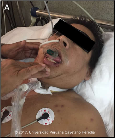
Epidemiology: Born and raised in Moyobamba, high rainforested area of San Martin Department, where he works as a farmer. No known TB exposure or high-risk behaviors for HIV. No known animal bites. Unknown vaccination history. Physical Examination: BP 120/90, HR 142, RR 32, T afebrile, SO2 95% (room air). Acutely ill appearance. Chest: clear to auscultation bilaterally. CV: tachycardic, no murmurs. Abdomen: distended, bowel sounds present, nontender no hepatosplenomegaly. Neurologic: Glasgow 15. Normal motor tone. No motor deficit, no meningeal signs. Muscle strength and deep tendon reflexes are preserved. Spasm of masseter muscles [Image A]. Laboratory Examination (on admission): Hct 39%. WBC 7.9 (no differential), platelets 280 000. INR 1.07. Glucose 166 mg/dL, BUN 20 mg/dL, creatinine 1.1 mg/dL. Normal electrolytes: Na 145 mEq/L, K 4.76 mEq/L, Cl 107 mEq/L, Ca 9.7 mEq/L, Mg 2.1 mEq/L. LDH 625 U/L (N<250), CPK 532 U/L (N < 170). sO2 99.8%, HCO3 18.5 mmol/L, lactate 0.9 mmol/L.
UPCH Case Editors: Carlos Seas, Clinical Course Coordinator/Sofia Zavala, Associate Coordinator UAB Case Editor: David O. Freedman, Course Director Emeritus |
|
Diagnosis: Generalized Tetanus
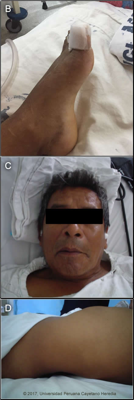
Discussion: On detailed examination a wound of the left great toe was present [Image B] and the patient admitted to an accident 12 days earlier involving a dirty axe. Generalized tetanus is a purely clinical diagnosis with highly characteristic features, and, as in this case, the diagnosis is usually made within minutes of arrival at a medical facility. In general, disease begins with trismus or lockjaw, which are spasms of the masseter muscles, although, initial symptoms may occur in other muscle groups as in the patient where the neck muscles were initially involved. After a variable period, the symptoms progress to generalized muscular rigidity, on which is superimposed increasingly severe generalized reflex muscular spasms manifested by the characteristic sardonic smile (risus sardonicus), opisthotonos (arched back) [Images C,D], and spasm of respiratory muscles and larynx. See also previous cases ( 2011-10, 2009-8). In severe cases there are prolonged spasms occurring less than 1 hour apart, and in very severe cases there is autonomic hyperactivity with sweating, fever, tachycardia, salivation, arrhythmias, hyper- or hypotension, hyperthermia, etc. Some aspects of generalized disease can be mimicked by hypocalcemic tetany, phenothiazine induced dystonia, epilepsy, rabies, strychnine poisoning, or narcotic withdrawal, but the history of wound (not elicited in up to 20%), epidemiology, and clinical course of tetanus usually lead to little confusion. Mild localized tetanus in which trismus does not progress to generalized disease with reflex spasms is rare. In the initial phase, the trismus itself has a broader differential diagnosis including dipththeria, parotitis, odontogenic abscess, peritonsillar abscess, retropharyngeal abscess, and traumatic injury, but in these cases trismus is due to pain and there is usually fever, whereas in tetanus trismus is due to masseter rigidity and initially there is no fever. Disease is caused by a toxin, tetanospasmin, released by Clostridium tetani that infect the wound. Spread of toxin is both retrograde through the affected axons as well as via blood to nerve endings in other parts of the body. Masseters are usually affected first due to their short axons. The action is pre-synaptic, irreversible, and blocks inhibitory neurotransmitter action leading to muscle spasm. Poor prognostic indicators include short incubation period (< 7 days) from time of the wound to onset of symptoms (12 days here), short period of onset (?? 48 hours) from onset of symptoms to first reflex spasm, and high-risk portal of entry (compound fracture, gynecologic, postoperative, intramuscular injections, and burns). Management is complex and must be done in a well-equipped Intensive Care Unit (ICU). Non-ICU care in severe cases is associated with high mortality. General principles are listed here, but detailed written dosing protocols must be available and used for most interventions. |