Key Insights
- UAB researcher Kenneth Chiou, Ph.D., contributed to the currently the most up-to-date and detailed multi-species cellular atlas of the basal ganglia, providing a foundational resource for future neuroscience research.
- Researchers have discovered that aging is dramatically different across cell types, brain regions and sexes, which can provide insights about risk for earlier onset disease.
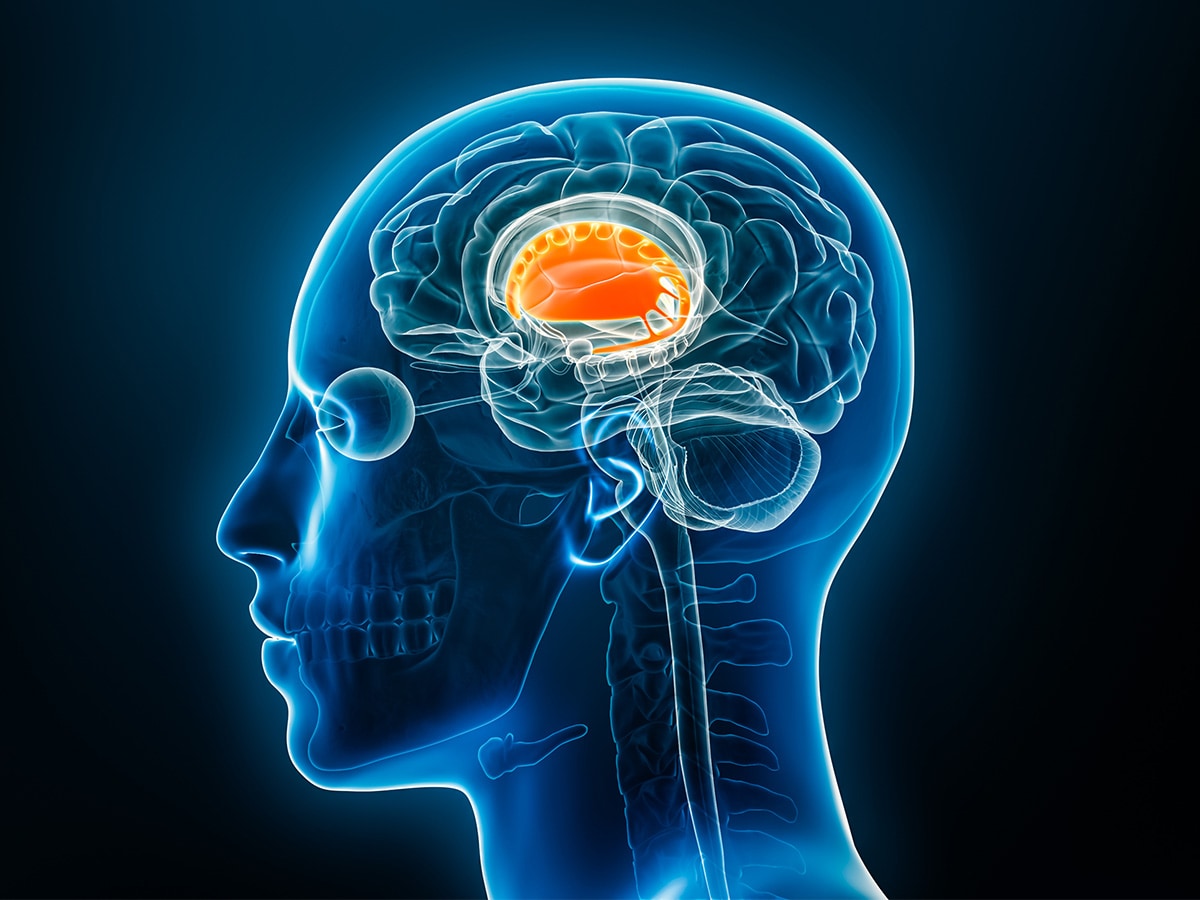 Researchers have discovered that aging is dramatically different across cell types, brain regions and sexes.Scientists and researchers across the world are working to better understand brain cells and aging’s impact on the brain. Dozens of labs and hundreds of scientists are part of a global effort to display their findings in a one-of-a-kind integrative atlas of the human basal ganglia.
Kenneth Chiou, Ph.D., assistant professor in the University of Alabama at Birmingham College of Arts and Sciences Department Biology, is among the leaders of a research team whose study is one of 28 articles featured in the package that explores brain aging discoveries.
The team’s findings in “Single cell, multi region profiling of the macaque brain across the lifespan” explores how brain cells age in a brain-region-dependent manner and are packaged in the Cell Press family of journals.
The research was co-led by a collaborative team including Noah Snyder-Mackler, Ph.D., of Arizona State University; Jay Shendure, Ph.D., M.D., of the University of Washington; Michael Platt, Ph.D., of the University of Pennsylvania; and Chiou. The team also includes co-first authors Wei Yang, UW, and Kelsi Watkins, ASU.
According to Chiou, “the Cell package represents the largest-yet effort to map out a reference of the human basal ganglia. Much in the same way that the Human Genome Project mapped out the human genome, which massively accelerated research into human genomic diversity, this team of neuroscientists pooled tremendous resources to map out a key region of the brain.”
Human basal ganglia are the structures in the brain that coordinate voluntary movement, habit formation, reward processing and decision-making.
The research team contributed to this effort by profiling the diversity of brain cell types across brain regions and among many individuals of a population. By focusing on a representative slice of the population, the team was able to characterize how cells change with age.
“We all have experiences with changed behavior, learning, memory and so on as we age,” Chiou said. When we dig deep into the brain, those changes can be explained in part by changes in how our brain cells function, which includes the genes that cells ‘express.’”
Do all parts of the brain age the same?
Through the team’s analysis, the team discovered that brain cells are not all impacted by aging the same; some cells are more vulnerable to aging than others.
The team’s study, published in Cell, provides new resources for aging research with the analysis of 5.3 million brain cells sampled from 11 brain regions. From these cells, the team identified 225 distinct subtypes of cells grouped into 12 broader cell classes.
 Kenneth Chiou, Ph.D.This study stands out among others in its area due to the scope and magnitude of this sample size.
What cells are vulnerable to aging?
The brain is the primary organ of the nervous system, and the two major cell types in the nervous system are glial cells and neurons. Support cells and non-neuronal cells like oligodendrocytes, glia and microglia are impacted differently by aging than neurons.
At a single-cell resolution, “the study identified age-depleted and vulnerable neuronal populations and subtle, age-associated compositional differences in glia,” according to the publication. Including cell type and age, the study revealed “sex-specific signatures that could underline biases in neurological disorders.” These insights also revealed that different areas of the brain do not age the same, which supports why certain abilities decrease as you age.
The team then merged these findings with transcriptional analyses of Alzheimer’s disease to gain insights in relations between normal and degenerative gaining.
As you get older, your risk of neurodegenerative diseases such as Alzheimer’s disease and Parkinson’s disease increase.
“By the time many clinical hallmarks of disease are detected, clinical options are limited, as many hidden cellular and molecular changes that have already occurred are irreversible,” Chiou said.
“Our study suggests that healthy aging among individuals who do not show evidence of disease shares many of the same molecular characteristics of disease, offering hope that individuals at risk for earlier onset disease may be identified and treated preventatively, guided by knowledge of the molecular characteristics of aging.”
What does this atlas mean for science?
“I hope readers grasp the sheer complexity of cell types and cell states,” Chiou said. “The deeper we look, the more we discover nuanced differences among cell types between brain regions and across individuals that, though small and difficult to detect, play an outsized role in explaining differences in how our brains function and how we age.”
While much about the relationship between brain aging and disease remains unknown, this article and the findings that make up the cellular atlas continue to pave the way for discoveries that could reveal the unknown of aging and health. According to Chiou, “This establishes crucial foundational knowledge upon which future generations of neuroscientists may benefit, including those documenting aging and neurodegeneration of the brain.”
The atlas is currently the most up-to-date and detailed multi-species cellular atlas and is the first in the continuing work of the BRAIN Initiative Cell Atlas Network. This achievement plays a large role in BICAN’s goal to develop an atlas of the cell types of the human brain.
Chiou’s work from this atlas is funded in part by UAB award R00 AG075241.