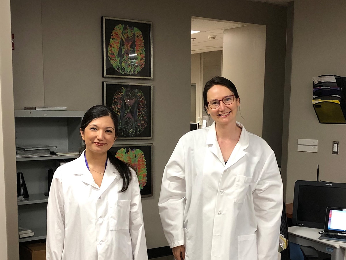
 Pinar Demirayak, Ph.D., and Kristina Visscher, Ph.D A new study from the University of Alabama at Birmingham suggests that, while each brain is wired differently, the portions used for demanding, attention-heavy tasks may become the most distinctly personal. The study, led by Pinar Demirayak, Ph.D., a researcher in the UAB Marnix E. Heersink School of Medicine, explores the idea that experience reshapes neural connections, making the brain’s networks better suited to their lived environment.
Rewriting the rules of sight
Researchers from the lab of Kristina Visscher, Ph.D., an associate professor in UAB’s Department of Neurobiology, found that the more attention a brain region receives, the more its connections to the rest of the brain become distinct across individuals, while regions used more passively look strikingly familiar from person to person.
To test this idea, Demirayak and her colleagues took advantage of a unique property of the visual system. Central vision, or the sharp detailed vision at the center of a person’s gaze, and peripheral vision, or vision outside of the direct point of gaze, are processed by separate regions of the brain’s primary visual cortex.
“We examined this in two contexts where different parts of primary visual cortex are used for more, or less, attention-demanding visual tasks,” Demirayak said. “We rely on the amazing fact that the visual cortex is retinotopically organized; that is, central vision is processed by different cortical regions than peripheral vision.”
Individuals with healthy eyesight rely heavily on central vision via the eye’s fovea for effortful visual tasks such as reading a page or scanning a face, while peripheral vision is rarely used for effortful tasks. Some individuals who lose their central vision, often due to macular degeneration, adapt by developing a preferred retinal locus, or an area of remaining peripheral retina that effectively becomes a substitute fovea.
Over time, this area becomes the primary region used for tasks that once relied on central vision, while the rest of the peripheral vision continues to function as normal. To further explore this occurrence, Demirayak zeroed in on a well-understood feature of the visual system that allowed researchers to compare heavily and lightly used regions of the brain side by side.
Fingerprints in the brain
Using scans from 23 individuals with healthy vision and 21 individuals with central vision loss, collected while they were at rest, Demirayak mapped how each region’s whole-brain connectivity pattern compared in each person. In the individuals with healthy vision, the results showed that the parts of the brain that process peripheral vision looked almost identical from person to person, while the parts of the brain that process central vision were highly individualized.
“We find that the whole-brain patterns of connections to the cortical representations of peripheral vision are quite similar from person to person,” Demirayak said. “On the other hand, when you look at whole-brain patterns of connections to central representations, they are idiosyncratic, like people’s fingertips.”
One exception to the rule
The second half of the study focused on individuals who had lost their central vision as adults. Their brains did not rewire dramatically — with one exception, in the exact location Demirayak’s theory would predict.
“We find that whole-brain patterns of connections of the visual cortex do not change much for people with central vision loss, except in the cortical representations of the preferred retinal locus, which people use preferentially for attention-demanding tasks.”
Because these patients developed vision loss later in life, the individualized pattern in their preferred retinal locus could not be attributed to patterns established early in brain development. Instead, it appeared to be a product of the specific way each person came to use that section of the cortex.
“The cortical representation of the preferred retinal locus shows more idiosyncratic patterns of connections than the central region,” Demirayak said. “This suggests that the idiosyncratic connections are associated with experience, not development, since these patients developed vision loss in adulthood.”
A new lens on plasticity
Beyond the visual system findings, the study proposes a broader methodological shift in how scientists study brain plasticity. Rather than comparing the average brain pattern of one group to another, the researchers measured how far each individual’s pattern strayed from the norm.
“We’re excited about the findings, in part, because it has opened our eyes to new ways of thinking about adult plasticity,” Demirayak said. “The field often examines neural plasticity by comparing the mean of one group to the mean of a control group.”
“That ignores the knowledge that each person’s pattern of brain connections starts out distinct, and an experience is likely to perturb each system slightly differently,” Demirayak said. “By examining the idiosyncrasy of the connections, we can identify plasticity in connection patterns.”
The researchers note the approach could eventually support more personalized rehabilitation strategies for people with vision loss, because a “one-size-fits-all” model can miss how differently each patient’s brain has adapted. More broadly, they suggest the same idiosyncrasy-based method could be applied to other kinds of experience-driven brain change, from the motor cortex of musicians to the brains of people who have lost limbs.
What emerges is a new picture of adult plasticity, not a dramatic rewiring, but a slow, individualized recalibration.
Additional authors of the study, published in the Journal of Neuroscience and titled “Increased Attentive Use Is Linked to More Idiosyncratic Functional Connections,” were lab members Pauline Stewart and Rachel Chua, and Leland Fleming, Ph.D., Texas A&M University.