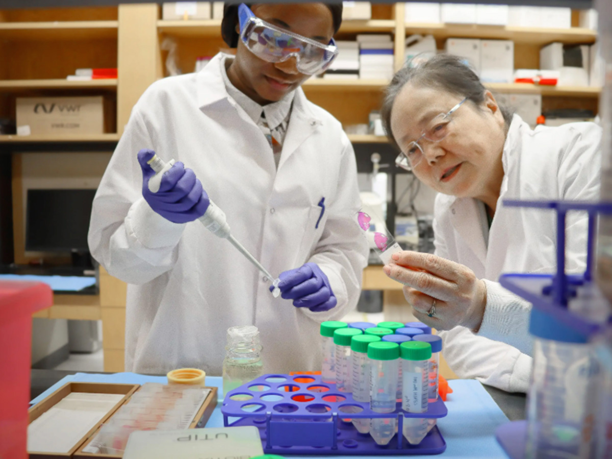
 UAB researcher and post-doctoral candidate, Sophia Bamishaye, Ph.D., with YangResearchers at the University of Alabama at Birmingham’s O’Neal Cancer Center are advancing a groundbreaking approach to cancer imaging that could one day allow physicians to identify and distinguish different types of cancer cells during a routine MRI scan. The innovative technology, being developed by Jenny Yang, Ph.D., combines precision molecular targeting with protein engineering to create MRI contrast agents capable of revealing the unique biological makeup of tumors.
Yang, an associate scientist at the O’Neal Cancer Center and a professor in the UAB Department of Medicine, has spent decades pioneering the development of protein-based imaging agents and biosensors. Internationally recognized for her work at the intersection of chemistry, molecular biology and biomedical engineering, Yang’s research focuses on designing proteins that can safely and precisely detect disease-related biomarkers within the body.
Her latest work centers on biomarker-targeting protein MRI contrast agents for precision MRI, a patented emerging technology that has the potential to fundamentally change how cancer is diagnosed and monitored over time.
Promising advancements in MRI screening
The study marks an important step toward next-generation molecular MRI. By targeting collagen, a key component of the tumor microenvironment that changes as cancer becomes invasive and spreads, Yang’s approach could help detect aggressive disease earlier and assess cancer progression more comprehensively than conventional imaging. The findings support the promise of her protein engineering approach for future clinical applications, including detecting cancers and metastases in the liver, pancreas, lung, colon, prostate and breast, as well as fibrosis in the liver, lungs, heart and kidneys.
A promising extension of this work is multicolor molecular MRI, which uses targeted contrast agents with distinct imaging signatures. This technology could allow clinicians to distinguish multiple cancer subtypes simultaneously, each displayed as a different color or signal, providing a more detailed picture of a tumor’s molecular diversity.
A traditional MRI provides detailed images of anatomy but offers limited insight into the molecular characteristics of cancer. Yang’s laboratory is engineering protein-based contrast agents that bind selectively to biomarkers on cancer cells. These agents produce distinct MRI signals that could reveal both a tumor’s location and the different cancer cell populations within it.
Visualizing tumor heterogeneity, the differences among cancer cells within a tumor, could help advance precision oncology. Tumors often contain distinct cell populations that respond differently to treatment, with some remaining sensitive while others develop resistance. Identifying these populations noninvasively could help physicians tailor therapies, monitor treatment response and potentially detect molecular changes before they become apparent on conventional imaging.
“Every patient’s cancer is biologically different,” Yang said. “Our goal is to give physicians the ability to see those molecular differences in real time without the need for repeated invasive biopsies. We envision MRI’s becoming a tool that not only shows where a tumor is located but also reveals the biology driving that cancer.”
Unlike many molecular imaging approaches that rely on radioactive tracers, Yang’s protein MRI contrast agents are designed for use with conventional MRI scanners already available in hospitals worldwide. The technology has the potential to provide comprehensive molecular information while allowing patients to undergo repeated imaging throughout treatment.
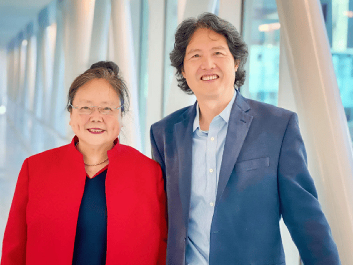 Jenny Yang, Ph.D., with Wei Zhou
Advancing toward human clinical trials
“Reaching this stage reflects years of collaborative scientific discovery,” Yang said. “Our next objective is to secure the resources needed to complete the regulatory pathway and bring this technology into human clinical trials, where we can begin evaluating its potential to improve cancer diagnosis and patient care.”
If successful, the technology could become one of the first protein-based molecular MRI platforms capable of simultaneously identifying multiple cancer biomarkers in patients. Such an advance would represent a significant step toward truly personalized imaging, enabling physicians to visualize the biological complexity of each patient’s cancer and tailor treatment accordingly.
Yang’s latest collaborative study, Early detection of invasive lung cancer and multiorgan metastasis by a collagen-targeted protein MRI contrast agent, was published in Science Advances, one of the world’s leading multidisciplinary scientific journals. Conducted in collaboration with Wei Zhou, M.D., of Emory University and Georgia State University, their research demonstrates the ability of a novel collagen-targeted protein MRI contrast agent to detect invasive lung cancer and identify metastatic disease in multiple organs in a single imaging examination.
“This is exactly the type of transformational science the UAB O’Neal Cancer Center is committed to advancing,” said Barry Sleckman, M.D., director of the O’Neal Cancer Center. “Dr. Yang’s research brings together exceptional innovation in chemistry, molecular imaging and cancer biology, with a clear focus on improving patient care. The opportunity to move this technology toward human clinical trials represents an exciting milestone with tremendous potential for precision oncology.”
Decades of innovation leading to a translational breakthrough
Yang has dedicated much of her scientific career to designing novel protein technologies that improve the diagnosis and treatment of disease. Her laboratory has developed numerous protein engineering platforms, molecular imaging probes and biomarker-targeting technologies, which have applications in cancer, cardiovascular disease and neurological disorders.
Yang’s multidisciplinary expertise in chemistry, protein engineering and molecular imaging has enabled collaborations with scientists and clinicians across the country, helping bridge the gap between fundamental laboratory discoveries and clinical applications. At the UAB O’Neal Cancer Center, Yang works alongside physician-scientists to ensure that new imaging technologies are developed with real patient needs in mind.
Yang is a 2025 Blazer Bridge Fund awardee, a program that supports UAB inventors in advancing early‑stage technologies, launched in 2023 by the Harbert Institute for Innovation and Entrepreneurship. Through utilizing these resources, Yang is further developing her novel Gd³⁺‑free, protein‑based MRI contrast agents, helping move the invention closer to future clinical translation.
Yang’s research led her to be selected for the Torsten Almén Award for Pioneering Research in Contrast Media presented by the Contrast Media Research symposium series. The award celebrates researchers whose work represents a significant and sustained contribution to the development of contrast media with the potential to advance the field of medical imaging.