 Long-held dogma says lung fibrosis in diseases like idiopathic pulmonary fibrosis, or IPF, results from recurrent injury to alveolar epithelium that is followed by dysregulated repair. Research at the University of Alabama at Birmingham uproots that paradigm, and it suggests a possible treatment target for IPF.
A. Brent Carter, M.D., and colleagues report in the Journal of Clinical Investigation that the recruited monocyte-derived macrophages, which have an increased flux in the mevalonate metabolic pathway — without any experimental injury — can induce lung fibrosis in a mouse model. When there is prior lung injury, the increased flux through the mevalonate pathway exacerbates the resulting fibrosis. The mechanism polarizes macrophages to a profibrotic state that causes pathogenic macrophage/fibroblast signaling.
Furthermore, study of humans with IPF showed that three hallmarks of the mechanism that leads to lung fibrosis in the absence of injury in mice are also found in bronchoalveolar, or BAL, cells from these patients, as compared to healthy individuals. The three are 1) activation of the small GTPase protein Rac1 and its localization into the intermembrane space of mitochondria in the BAL cells, 2) increased production of mitochondrial reactive oxygen species by BAL cells from patients with IPF, and 3) evidence of increased flux through the non-sterol arm of the mevalonate pathway in the BAL cells results in the augmented activation of Rac1.
“Here, we show a paradigm shift that indicates a critical and essential role for monocyte-derived macrophage/fibroblast crosstalk in the development and progression of fibrosis in the absence of epithelial injury,” said Carter, a professor in the Division of Pulmonary, Allergy and Critical Care Medicine, UAB Department of Medicine. “Although alveolar epithelial cell dysfunction may initiate the development of IPF, our findings suggest that repeated epithelial cell injury is not required for the progression of fibrosis.”
“We propose that monocyte-derived macrophage/fibroblast crosstalk can induce fibrosis without epithelial cell injury and is the primary driver in mediating disease progression,” Carter said. “These observations suggest that targeting the mevalonate pathway may abrogate the role of macrophages in dysregulated fibrotic repair.”
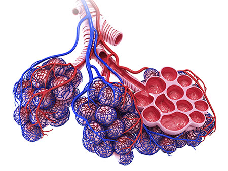
Macrophages are large white blood cells that are the initial defense-fighting cells in the lung. In 2016, Carter and colleagues showed that lung macrophages are the primary source of the damage-inducing TGF-β1 — a potent cytokine — in the lung.
Pulmonary fibrosis is a progressive disease with irreversible scarring of the lungs. IPF, one form of pulmonary fibrosis, has a median life expectancy of three to five years after diagnosis.
 A. Brent Carter, M.D.Study details
The mechanism for lung damage without injury that Carter’s team found has the following steps:
In macrophages, an increased flux through the non-sterol arm of the mevalonate pathway increased the production of geranylgeranyl diphosphate, or GGDP. This increased flux is secondary to greater levels of acetyl-CoA from metabolic reprogramming of the macrophages to β oxidation.
Using GGDP, an enzyme then adds geranylgeranyl, a lipophilic membrane anchor, to Rac1, a Rho GTPase, and the geranylgeranylated Rac1 translocates from the cytosol to the intermembrane space of mitochondria. There, it induces production of reactive oxygen species.
The reactive oxygen species cause a phenotypic shift of the macrophage to a profibrotic state, acting through key transcription factors. The profibrotic macrophages then produce TGF-β1, which transforms fibroblasts into pathogenic myofibroblasts.
The study, “Increased flux through the mevalonate pathway mediates fibrotic repair without injury,” is published in the Journal of Clinical Investigation. Co-authors with Carter are Jennifer L. Larson-Casey, Mudit Vaid, Linlin Gu, Chao He, Guo-Qiang Cai, Qiang Ding, Dana Davis, Ranu Surolia and Veena B. Antony, UAB Department of Medicine, Division of Pulmonary, Allergy, and Critical Care Medicine; Taylor F. Berryhill, Landon S. Wilson and Stephen Barnes, UAB Targeted Metabolomics and Proteomics Laboratory; Jeffrey D. Neighbors and Raymond J. Hohl, Pennsylvania State University; Kurt A. Zimmerman and Bradley K. Yoder, UAB Department of Cell, Developmental and Integrative Biology; and Ana Leda F. Longhini and Vidya Sagar Hanumanthu, UAB Department of Medicine, Division of Clinical Immunology and Rheumatology.
Support came from National Institutes of Health grants ES015981-12 and ES027464-01, Department of Veteran Affairs grant CX001715-01, a Parker B. Francis fellowship, and American Lung Association grant RG-507440.
At UAB, Carter holds the Department of Medicine Endowed Professorship in Lung Immunology, and he is scientific director of the UAB Interstitial Lung Disease Program.