 |
Gorgas Case 2001-09 |
 |
|
The following case was seen on the ward of the Tropical Medicine Institute of Cayetano Heredia Hospital in Lima by the 2001 Gorgas Course participants.
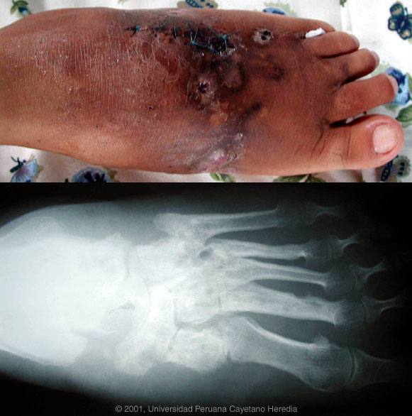  History: 35 yo female with a 10 year history of a slow growing swelling on the left foot associated with mild pain. Gradual onset of deformity. More recently draining fistula with secretions containing white grains has been noted. No fever or systemic symptoms. Patient still able to walk. Past history and review of systems is non-contributory. Epidemiology: Born and lives in Huanta in the Ayacucho Department, a rainforest area. PE: Afebrile. Findings restricted to the deformity of the left foot (shown in photograph) and the draining sinus tracts. Labs/X-ray: Normal WBC. Normal CXR, liver function. X-ray of the affected foot (see photo) shows numerous lytic lesions with massive destruction of the cuneiforms, navicular, calcaneous and involvement of all the proximal phalanges.
|
|
Diagnosis: Actinomycetoma with osteomyelitis.
Discussion: A deep incisional biopsy of soft tissue and bone was performed. Histopathology demonstrated acute and chronic inflammatory reaction and small grains typical of actinomycetoma. A silver stain demonstrated very thin filamentous bacteria and gram stain of the grains showed gram positive bacteria, both of which are compatible with actinomycetoma. Actinomycetoma refers to a mycetoma with a bacterial cause usually due to Nocardia, Streptomyces (white/yellow grains), or Actinomadura (red grains). The main and important differential diagnosis in this case is eumycetoma, a mycetoma due to fungal causes (Madura foot) [see Gorgas Case 2002-04]. This common subcutaneous mycosis in the tropics is caused by a wide variety of fungal organisms but is usually due to Madurella mycetomatis. In contrast to bacterial actinomycetomas, which are responsive to antibacterial agents such as TMP/SMX + Aminoglycoside or Rifampicin or Dapsone, eumycetomas are poorly responsive to medical therapy and usually require aggressive surgery or amputation. Clinical diagnosis is thus not sufficient in these cases and an exact agent must be ascertained if possible. Other diagnoses in the differential include TB, botrymycosis (chronic staphylococcal infection) [see Gorgas Case 2003-04], other causes of chronic bacterial osteomyelitis, and malignancy. Other subcutaneous mycoses include sporotrichosis, chromomycosis and lobomycosis (phaeohyphomycosis). Eumycetoma, and in this case bacterial infection due to actinomycetoma, appear to be localized phenomena due to local innoculation without any potential for widespread dissemination.
|