|
Diagnosis: furuncular myiasis caused by Dermatobia hominis
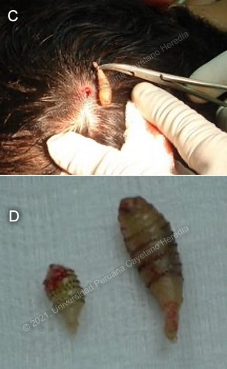
Discussion: Surgical removal of the maggots was performed successfully, one immature larvae of the classical human botfly Dermatobia hominis was obtained from each of the two lesions (Images C and D).
Myiasis is defined as the human infestation or other vertebrates with larvae (maggots) of Diptera (or true flies), with the larvae feeding on the host´s living or necrotic tissue. Myiasis can be classified based on anatomical parameters or in ecological terms. Clinicians prefer the anatomical classification: bloodsucking, cutaneous, wound and cavitary myiasis [Clin Microbiol Revs 2012;25:79]. The families of Diptera causing various forms of myiasis are very diverse and many have specific geographic distributions.
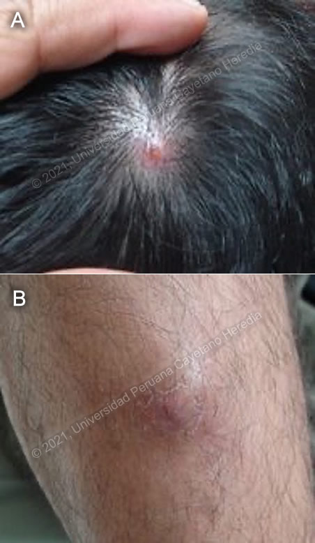
In furuncular myiasis (cutaneous myiasis) the larva penetrates intact living skin but cannot penetrates beyond the subcutaneous tissue developing into an expanding boil-like lesion, feeding on living tissues. The full life cycle may take up to 60 days, during this time the larva maturates from as small as 6mm length to reach a maximum of approximately 2cm. When not removed before, the third stage larva comes out and pupates on soil [J Clin Aesthet Dermatol. 2013;6:47-9]. In the tropical Americas, furuncular myiasis is due to Dermatobia hominis, the human botfly. The adult female fly physically captures a mosquito and then glues her eggs to the abdomen of the carrier, when the mosquitos feeds from humans a larvae inside burrows through the skin. Botfly lesions are almost always single, and the point of the furuncle is characterized by a small punctum through which the larva must obtain oxygen [see case #6, 2005]. Discharge of an exudative material or air bubbles through the small punctum is characteristic and should raise suspicion of furuncular myiasis. Some patients mention that they feel the larva moving inside the skin, our patient did not have that feeling. Human exposure to the fly habitat as a result of tourism or business activities has increased the recognition of this infestation [Clin Microbiol Revs 2012;25:79]. Furuncular myasis accounted for 6% of all dermatological problems among returned Israeli travelers in a study; 80% of these cases acquired the infestation in South America and most of them required manual extraction only (76%). Bacterial superinfections were extremely rare (1%) [J Trav Med 2015;22:232]. The differential diagnosis of furuncular myiasis includes pyodermitis, tungiasis and insect bites. Tungiasis results from the penetration of the skin by the flea Tunga penetrans, the lesions are located usually on the feet, suspect under the presence of painful, pruriginous lesions in nail folds, toe webs, or tips of toes.
Occlusion of the respiratory punctum such as with paraffin, petroleum jelly, adhesive tape or any occlusive dressing often causes the pear-shaped larva to come out and is curative. Manual removal is indicated when the maggot does not come out after occlusion and can be performed by squeezing the larvae, use of forceps or injecting 2ml of lidocaine underneath the nodule to press the larvae to come out to the surface. [J Am Acad Dermatol. 2008;58:907-26]. This procedure is challenging as the maggot has spines in its surface that attaches to the skin and in addition, the head of it is wider and located deep in the skin tissue. Surgical excision under local anesthesia is necessary when occlusive and manual extraction fail to remove the maggot (refractory cases). A cruciate incision around the central punctum is recommended, irrigation and debridement may be needed to remove any residual tissue that may induce a severe inflammatory reaction later [J Clin Aesthet Dermatol. 2013;6:47-9].
Professor Hugo Lumbreras, the founder of our Tropical Medicine Institute, used to teach of the traditional approach to myiasis in Peru, which involved the occlusion of the entrance of the wound by a solution made from basil leaves, the odor of which forced the larvae out of the wound. The patient rejected the idea of suffocating the maggots or manually extracting them and requested a surgical extraction right away.
In Africa, furuncular myiasis is caused by Cordylobia anthropophaga or Tumbu fly (not to be confused with tungiasis). The Tumbu fly directly lays her eggs on dirty or soiled clothing. If not properly washed and ironed (heat kills the larvae) the eggs hatch and invade skin when the clothing is next worn [Am. J. Trop. Med. Hyg 2020;102: 251]. Lesions are almost always numerous and tend to be angrier and more erythematous than with the botfly. Bacterial superinfection is extremely uncommon with Cordylobia anthropophaga.
|