Explore UAB

University of Alabama at Birmingham

Gorgas Case 2023-10

Universidad Peruana Cayetano Heredia
The following patient was seen as an inpatient in the Internal Medicine ward of Cayetano Heredia Hospital in Lima by the 2023 Gorgas Advanced Course participants.
Image for Case 2023-10

History: A 54-year-old patient presented with a one-week history of intermittent dizziness and holocranial headaches. On the day of admission, he presented sudden-onset weakness and numbness in the upper right extremity and decided to go to the emergency room. On the way there, his family describes an episode of transient loss of consciousness with sphincter relaxation and a post-ictal period.

Epidemiology: Born in Cajamarca, in the northern highlands of Peru, where he lived until 8 years of age in a farm in a rural community and cared for animals including cows, pigs, sheep and dogs. Lived in Trujillo until 18 years of age, working in animal husbandry. Currently lives in the city of Lima, where he has no pets. Traveled back to Cajamarca one year before presentation.

Past medical history: Episode of Bell’s palsy in 2019, which resolved after treatment with steroids. No known tuberculosis contacts.

Physical Examination:
 HR 86, RR 16, T 37, BP 140/70, SatO2 98% (FiO2 0.21). Neuro: Alert and oriented x3, Glasgow Comma Scale 15/15. Motor strength 4/5 in right upper extremity, rest 5/5. No sensory deficits. No nuchal rigidity. Rest non-contributory.

Laboratory: Hb 15, Plt 290 000, WBC 17 960 (0% bands, 0.11% eosinophils, 16.6% neutrophils, 0.5% lymphocytes). Glucose 120, Na 146, K 4.08, Cl 108.

Imaging: Brain CT is shown in Images A1, A2. A right lower limb X-ray is shown in Image B.


UPCH Case Editors: Carlos Seas, Course  Director / Paloma Carcamo, Associate Coordinator

UAB Case Editors: David O. Freedman, Course Director Emeritus / German Henostroza, Course Director

{slide=CLICK HERE FOR DIAGNOSIS & DISCUSSION OF THE CASE ABOVE

Diagnosis: Parenchymal neurocysticercosis

Images for Case 2023-10
image overlay

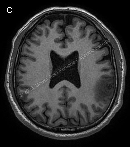
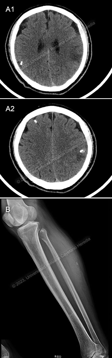
Discussion: Brain CT showed a calcified lesion with surrounding edema in the left parietal lobe and additional calcified nodular lesions with no edema in the left occipital lobe. The lower limb x-ray showed a cigar-shaped calcification compatible with a calcified cysticercus. Brain MRI (Image C) confirmed these findings, showing an irregular nodular cortico-subcortical lesion of heterogenous aspect with peripheral vasogenic edema in the left parietal lobe, and the previously described left occipital lesions. A diagnosis of neurocysticercosis was made with these images.

Neurocysticercosis is caused by brain tissue invasion by the larval stage of Taenia solium. The helminth maintains its life cycle between humans and pigs. Humans, the definitive hosts for T. solium, acquire intestinal taeniasis after ingestion of cysticerci in undercooked pork meat, the usual intermediate host. Humans can become accidental intermediate hosts following ingestion of human feces with eggs and gravid proglottids; self-infection can also happen. After ingestion, oncospheres hatch into the intestine, invade the intestinal wall, and enter the bloodstream to invade different organs where they mature into cysticerci.

T. solium has complex mechanisms to avoid the host immune response; thus, initially cysts do not cause much local inflammation or symptoms. After some time, the immune system starts to recognize the parasite and edema appears surrounding the cysticerci, such as was seen in our patients imaging. Eventually, cysts begin to degenerate and eventually become calcified granulomas. The calcified lesions in our patient’s brain imaging indicate that he was likely infected many years ago and has been asymptomatic until his current presentation.

Symptoms of neurocysticercosis depend on the location and stage of the cysticerci. They are most commonly intraparenchymal, and usually lead to focal seizures which may generalize. Unusually, an extremely high parasite burden can lead to an encephalitis-like picture caused by diffuse cerebral edema. Many cases are asymptomatic and identified incidentally on imaging. The parasite can also invade the cerebral ventricles, the subarachnoid space, the spine or the eye; these extra parenchymal cysts lead to varied clinical pictures.

A diagnosis of neurocysticercosis can usually be established with careful history-taking and neuroimaging. CT scans are more sensitive for detection of calcified lesions, while MRIs allow for better observation of cystic lesions. Observation of a scolex inside a cystic lesion in a brain MRI confirms the diagnosis. If neuroimaging is not confirmatory, serologic testing with enzyme-linked immunotransfer blot (EITB) can be performed, though sensitivity may be poor in patients with a single parenchymal lesion or only calcified lesions. ELISAs are not recommended as they have high rates of false positives and negatives1.

Treatment for neurocysticercosis should initially focus on management of seizures and any other complications. Antiepileptic treatment should be started promptly and continued until resolution of cystic lesions on brain imaging and a significant time without seizures. The World Health Organization recommends tapering off antiepileptics 6 months after the last seizure in individuals with a single enhancing lesion and low risk of seizure recurrent, but continuing antiepileptics for at least 2 years in people with single or multiple calcified lesions and epilepsy2. IDSA and ASTMH recommend tapering off antiepileptic drugs after resolution of cystic lesions and no seizures for 24 consecutive months1.

Anthelmintic treatment is not urgent but should be administered to all patients with viable or degenerating cysts, except in cases of untreated hydrocephalus or cysticercal encephalitis. Anthelmintic treatment correlates to better outcomes in terms of cyst resolution and seizure control, and may hasten radiologic resolution of the lesions2. For patients with 1-2 viable parenchymal cysts, treatment should be given with albendazole monotherapy. Albendazole can be combined with praziquantel for patients with more than 2 viable cysts. Anthelmintic treatment may lead to perilesional edema and exacerbation of symptoms, thus concomitant administration of corticosteroids recommended and has been shown to be effective in reducing edema and subsequent seizures and lesion calcification. After antiparasitic treatment, neuroimaging should be repeated at least every 6 months to confirm resolution of cystic lesions. IDSA guidelines do not recommend anthelmintic treatment for patients with only calcified parenchymal lesions; these patients seem to benefit mostly from symptomatic therapy1.

Our patient received acute antiepileptic treatment with diazepam upon admission and was started on phenytoin. He will receive treatment with albendazole 15 mg/kg/day for 14 days with adjunctive corticosteroids because of the single inflammatory lesion on MRI and will be monitored as an outpatient for radiological resolution of the cystic lesion and seizure control.

References

1. White AC Jr, Coyle CM, Rajshekhar V, et al. Diagnosis and Treatment of Neurocysticercosis: 2017 Clinical Practice Guidelines by the Infectious Diseases Society of America (IDSA) and the American Society of Tropical Medicine and Hygiene (ASTMH). Clinical Infectious Diseases. 2018;66(8):e49-e75. doi:10.1093/cid/cix1084
2. World Health Organization. WHO guidelines on the management of Taenia solium neurocysticercosis. Published online 2021. https://www.who.int/publications/i/item/9789240032231

{/slide}