Explore UAB

University of Alabama at Birmingham

Gorgas Case 2023-04

Universidad Peruana Cayetano Heredia
The following patient was seen on the inpatient ward of Cayetano Heredia Hospital in Lima by the 2023 Gorgas Course participants.
Image for Case 2023-04

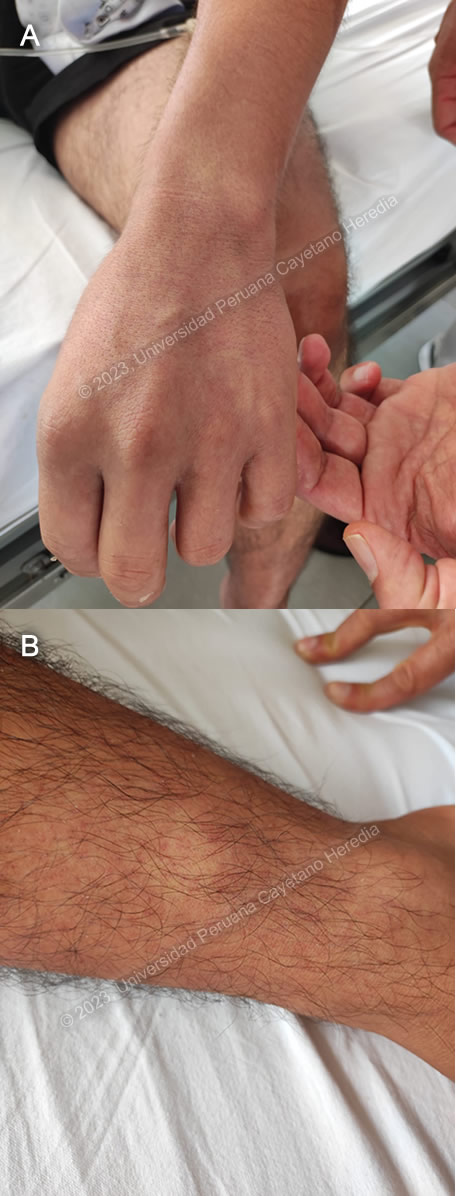
History: A 20-year-old previously healthy male patient presented with a 7-day history starting with high fevers (quantified at 39.5°C), headache, myalgias, arthralgias and retroocular pain. Three days before admission, he started to have nausea, one episode of vomiting and diarrhea (watery, no mucus or blood), and noticed a rash which started on his back and then generalized; the rest of the symptoms persisted. Two days before admission, the diarrhea and vomiting ceased but all other symptoms persisted. On the day before admission, he sought medical care and had blood tests drawn. He was referred to our institution with the results of these tests. He denied any bleeding.

Epidemiology: Born in Jaen, Cajamarca, in the northern high jungle of Peru, where he lived until he was 13 years. Currently lives in Lima, where he is a 2nd year medical student. He travels back to Jaen during school holidays to help with the family business, a fish import company. He was last in Jaen two months before admission, and he remained there until the day before admission when he traveled to Lima. He swam in rivers but not stagnant waters, did not go into caves, used insect repellent intermittently, and was bitten by mosquitoes multiple times. No animal exposures. No known tuberculosis exposures.

Past medical history: Had dengue at 8 years of age, while living in Jaen. Has received all childhood immunizations, including yellow fever vaccine. Drinks alcohol socially, no smoking or other recreational drug use. No sexual contacts in the last four months.

Physical Examination:
 BP 100/70 mmHg, HR 78 bpm, RR 17, T 37°C. Skin: confluent erythematous macular rash with islands of unaffected skin in arms, thorax, back and legs (Image A and B). Lymph: Bilateral femoral lymph nodes, 0.5mm in diameter, soft and non-tender. Abdomen: Non-tender, no visceromegaly. Rest of exam non-contributory.

Laboratory: Hemoglobin 15.5, hematocrit 47. WBC 5.27 (54.1% lymphocytes, 0% bands, 9.5% monocytes, 34.4% neutrophils, 1.8% eosinophils, 0.1% basophils). Platelets 46 000. PT 12.8, aPTT 44.4, INR 0.93. Glucose 83, Urea 30, Creatinine 0.53. AST 292, ALT 195, Alkaline phosphatase 103, GGT 224. HIV-1/2 negative.

Imaging: CXR was unremarkable. FAST ultrasound revealed scant liquid in pericardium and right pleural space, and no free liquid in the abdominal cavity. Gallbladder size was within normal limits, with a wall thickness of 2mm


UPCH Case Editors: Carlos Seas, Course  Director / Paloma Carcamo, Associate Coordinator

UAB Case Editors: David O. Freedman, Course Director Emeritus / German Henostroza, Course Director

{slide=CLICK HERE FOR DIAGNOSIS & DISCUSSION OF THE CASE ABOVE

Diagnosis: Presumptive dengue without alarm signs.

Images for Case 2023-04
image overlay

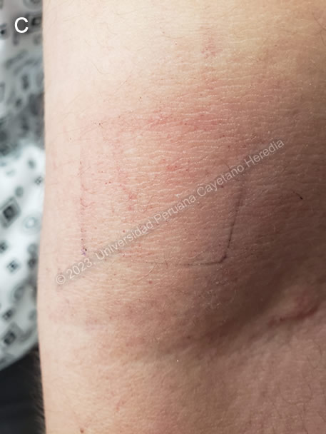
Discussion: A Thick and thin smears were negative for malaria, and a COVID-19 antigen test was negative. Though no confirmatory tests for dengue were performed, a presumptive diagnosis was made on the basis of the patient’s symptoms, exposure, and exclusion of other possible etiologies. During the first week of illness, a diagnosis of dengue can be established with NS1 (viral antigen nonstructural protein 1) detection or PCR, though sensitivity of NS1 diminishes in secondary infections1. ELISA for IgM can be positive from four days after symptom onset, thus this is the more appropriate test for this patient. Results are pending. A tourniquet test was performed by inflating a sphygmomanometer to the patient’s mean arterial blood pressure and keeping it inflated for 5 minutes. Afterwards, the number of petechiae in an area measuring one square inch in the antecubital fossa was counted (Image C). If there are more than 10 petechiae, as was the case with this patient, the tourniquet test is considered to be positive.

According to the World Health Organization (WHO) classification schemes published in 20092, a presumptive diagnosis of dengue can be made in a patient with history of travel or residence in an endemic area who presents with fever and at least two of the following: nausea or vomiting; rash; headache, eye pain, myalgias or arthralgias; leukopenia; and/or a positive tourniquet test. Additionally, WHO outlines seven warning signs of plasma leakage: abdominal pain, persistent vomiting, clinical fluid accumulation, mucosal bleeding, lethargy or restlessness, hepatomegaly >2 cm, or an increase in hematocrit concurrent with rapid decrease in platelets. Our patient presented with none of these signs, and thus did not warrant hospitalization.

Dengue usually presents with three phases of infection. In the initial febrile phase, patients present with high-grade fever, headaches, vomiting, myalgia, arthralgias, and occasionally a transient macular rash. After defervescence of fever, the critical phase begins where a small number of patients, particularly those with a second dengue infection, may develop plasma leakage, bleeding, organ impairment and shock. During this phase, ultrasound can be useful to rule out fluid accumulation in the pericardium, pleural space, or abdomen. A gallbladder wall over 3mm in thickness has been associated with more severe cases of dengue, while a thickness greater than 5mm may indicate a higher risk of hypovolemic shock3. Thrombocytopenia, as was seen with our patient, is common during this phase, and platelet counts usually rapidly improve during the recovery phase. The platelet count for the presented patient went up to 112 000 prior to discharge on day 2. During the recovery phase, the typical “red islands on a white sea” rash, as was seen in our patient (Image A and Image B), may appear.

This patient would have been a good candidate for vaccination with CYD-TDV (Dengvaxia), which is now available in Peru. It has been approved by the WHO for persons aged 9-45 years with confirmed previous dengue infection and living in endemic areas and protects especially against dengue infection requiring hospitalization4,5. However, because the vaccine increases risk of severe infection among children with no previous exposure to dengue, potentially eligible persons must be carefully evaluated prior to administering the vaccine. It is most effective against dengue serotypes 3 and 4 (approx. 75%); efficacy is lower against serotypes 1 (50%) and 2 (35-42%). All four serotypes are currently circulating in Peru. Additionally, logistics for vaccination can be challenging both for travelers and for persons living in endemic areas, as all three doses of the vaccine must be administered over a year.


References:
1. Ea H, J MJ, M B, et al. Performance of Dengue Diagnostic Tests in a Single-Specimen Diagnostic Algorithm. The Journal of infectious diseases. 2016;214(6). doi:10.1093/infdis/jiw103
2. World Health Organization. Dengue guidelines for diagnosis, treatment, prevention and control : new edition. Published online 2009. https://apps.who.int/iris/handle/10665/44188
3. Tavares M de A, João GAP, Bastos MS, et al. Clinical relevance of gallbladder wall thickening for dengue severity: A cross-sectional study. PLoS One. 2019;14(8):e0218939. doi:10.1371/journal.pone.0218939
4. Capeding MR, Tran NH, Hadinegoro SRS, et al. Clinical efficacy and safety of a novel tetravalent dengue vaccine in healthy children in Asia: a phase 3, randomised, observer-masked, placebo-controlled trial. Lancet. 2014;384(9951):1358-1365. doi:10.1016/S0140-6736(14)61060-6
5. Villar L, Dayan GH, Arredondo-García JL, et al. Efficacy of a tetravalent dengue vaccine in children in Latin America. N Engl J Med. 2015;372(2):113-123. doi:10.1056/NEJMoa1411037


{/slide}