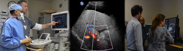
The abdominal imaging fellowship program at UAB provides 12 months of advanced training in computed tomography (CT), ultrasound (US), magnetic resonance imaging (MRI) and non-vascular interventional procedures of the abdomen and pelvis with state-of-the-art equipment.
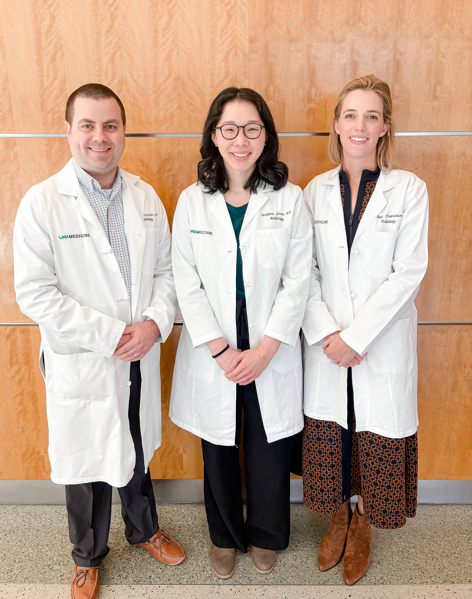
The program provides varied experiences that will serve as a solid foundation for either academic or private practice careers. The Abdominal Imaging section has faculty who provide coverage for body CT, US, MR, image-guided interventional procedures, and GI/GU fluoroscopic exams. Our abdominal imaging faculty members have a wide range of experience, accomplishments, and interests on the institutional, national and international levels. Several are widely known experts in abdominal imaging, with numerous scientific publications as well as prominent roles in national radiology organizations.
 Fellowship Experiences
- Learn to protocol and interpret complex cross-sectional abdominal and pelvic imaging studies using advanced imaging equipment and techniques
- Become proficient in performing CT and US-guided image-guided interventional procedures
- Have opportunities and mentoring to perform imaging research based on their personal goals
Body Computed Tomography
Approximately 150 – 200 abdominal and pelvic CT scans in a wide variety of clinical complexity are performed per day across all our inpatient and ambulatory facilities: University Hospital, The Kirklin Clinic of UAB Hospital, UAB Highlands, UAB Gardendale, and UAB Leeds.
Ultrasound
Approximately 140 – 150 ultrasounds are performed per day across all our facilities. Sonographer Radiologist Assistants assist with the section workflow.
A wide variety of sonograms are performed, including:
- US contrast agent evaluation, primarily of renal and hepatic lesions
- Liver elastography
- General abdominal (hepatobiliary and renal)
- Pelvis - female
- Small parts (thyroid, testis)
- Obstetric (1st trimester)
- Lower and upper extremity arterial and venous, carotids
- Preoperative mapping prior to hemodialysis access and postoperative hemodialysis AVF and graft evaluation
- Transplant - liver, renal, and pancreas ultrasound
- About 60-70% of the examinations are vascular
Magnetic Resonance Imaging
We perform approximately 30-50 MRI scans of the abdomen and pelvis across our facilities daily. UAB is one of the largest transplant and prostate cancer centers in the southeast.
Our fellows get extensive experience reading a variety of body MR scans, including:
- Complex hepatobiliary, pancreatic, gastrointestinal, and genitourinary cases
- Transplant imaging – Liver, pancreas, and kidneys
- Prostate imaging
- MR enterography
- Pelvic floor imaging
- MR elastography and fat/iron quantification
- MR imaging of the acute abdomen in pregnancy
Interventional Procedures
We perform a wide variety of non-vascular procedures in the abdominal imaging section. Fellows rotate through the interventional service with the option of additional elective interventional time if desired. We use ultrasound and CT guidance for biopsies and aspirations.
These procedures include:
- Liver, kidney, adrenal, lymph node, and other soft tissue and retroperitoneal biopsies
- Transplant kidney biopsies
- Thyroid biopsies
- Aspiration/drainage of thoracic, abdominal, and pelvic fluid collections
- Thoracentesis and paracentesis
- Renal and hepatic lesion microwave/radio-frequency ablations
Teaching and Clinical Conferences
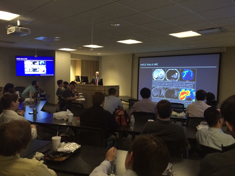
Viewbox teaching occurs daily and fellows often participate in teaching both residents and medical students. Though we do not require that our fellows give formal lectures to the residents, the opportunity to do so is an option. Fellows may also conduct board review sessions for senior residents.
Fellows can also attend any of several regularly scheduled interdepartmental clinical-radiologic case conferences. The conferences are held jointly with the divisions of gynecologic oncology, transplant surgery, gastroenterology, urology, surgical oncology, and GI surgery. These informative sessions are valuable opportunities for interacting with our referring clinicians.
Research
Fellows are encouraged, but not required, to become involved in projects during the fellowship year. Supervision and guidance are also available to help fellows initiate new projects. The fellows' research activities often lead to scientific articles and presentations at national meetings. Though most research conducted in the department is clinical, extensive resources exist for a broad variety of research endeavours. In addition to our clinical faculty, we have a large physics group, which includes the imaging informatics group. Extensive computer and statistical resources are also available.
Call Responsibilities, In-house Moonlighting, and Benefits
Routine clinical responsibilities are on weekdays only. Faculty and fellows share cross-sectional imaging call, with each person averaging about two weekends every six months. In some instances, fellows will instead occasionally take general radiology call, which primarily includes coverage for radiographic studies (chest, abdomen, and MSK).
Several moonlighting opportunities are available to our fellows, including potential opportunities both at UAB and in nearby community hospitals.
Salary, conference and vacation time, and fringe benefits are competitive with other fellowship programs. Fellows are on the same footing as faculty members in choosing vacation days, making vacation time generally flexible.
Birmingham City
Located in the heart of the southeast, Birmingham is a short drive from Atlanta, Nashville, Chattanooga, New Orleans, Memphis, and gulf coast beaches. Some of the most beautiful suburbs in the United States surround the city. With its picturesque surroundings, Birmingham has been recognized as an “All American City,” one of the top ten American cities in which to live and work, and one of the top ten entrepreneurial and job growth hot spots in America. The combination of pleasant weather, geography, activities, and lifestyle make this city a wonderful place to live, raise a family, and practice medicine.
General Information
How to apply
Thank you for your interest in the UAB Abdominal Imaging Fellowship Program. If you are interested in applying for our abdominal imaging fellowship, please submit the following:
- Application along with three letters of recommendation (one from program director)
- Personal statement
- Current curriculum vitae
- USMLE and medical school transcripts
Note: Three years of ACGME are required in the state of Alabama.
Leadership
 David Summerlin, M.D.