The Division of Molecular Imaging and Therapeutics program at The University of Alabama at Birmingham (UAB) is accredited by the Accreditation Council for Graduate Medical Education (ACGME). The fellowship is open to those applicants that have completed or will complete a residency in an ACGME accredited program.
Fellowship Overview
 The length of the Nuclear Medicine fellowship is determined by the residency completed by the applicant. For applicants from radiology residencies, the fellowship will last or one year. For applicants from internal medicine, pathology, or other non-radiology residencies, the fellowship will last for two years. The goal of the fellowship program is well-rounded experience in traditional nuclear medicine and molecular imaging. Applicants that complete this program can expect board eligibility for American Board of Nuclear Medicine certification, and eligibility for authorized user status.
Fellows in this program are actively involved in daily triage of patients to ensure that the appropriate study is performed, and that the best scintigraphic data obtainable is collected. The fellows in the program play a major role in the interpretation of the study's data and formulation of the Nuclear Medicine report. The fellows are involved in all teaching sessions, play a major role in diagnostic consultation, and first-and-foremost, are always conscious of the quality of patient care. The general Nuclear Medicine program includes all areas of scintigraphic imaging, radionuclide therapy, use of radionuclides in the ascertainment of laboratory physiologic values, and proper obtainment of in vivo tracer kinetic studies. Specifics regarding length of training and other requirements can be found below.
The research section contains two laboratories (one involved with new isotope development for evaluation of cancer and gene therapy, and a second using high field MRI to evaluate fMRI and MRSI protocols). There are four Ph.D. in the division of Nuclear Medicine who head these laboratories, and can assist you with developing and conduction experiments in these areas.
Instruction is provided through formal lectures, regularly scheduled conferences, and supervised clinical experience. Introductory and review materials are presented in structured training sessions. Structured didactic and laboratory physics instruction is provided by the physics department. Conference and journal club topics are selected to complement the curriculum.
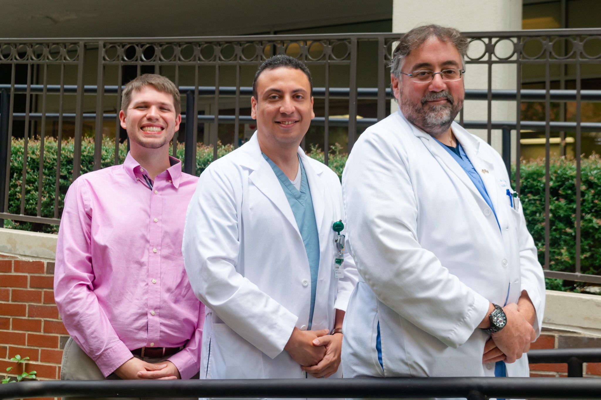
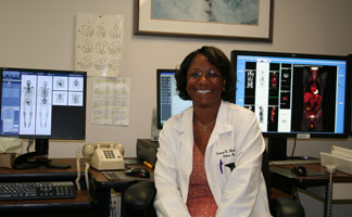
Molecular Imaging and Therapeutics fellows rotate through the Division of Molecular Imaging and Therapeutics at University Hospital, the Nuclear Medicine Service of the Veterans Affairs Medical Center at Birmingham (V.A.), and the Children's Hospital. These units function in close cooperation with one another. The staff of the Division of Molecular Imaging and Therapeutics includes eight physicians (two with M.D./Ph.D. degrees), two physicists, two research assistants, nine technologists, and four clerical personnel. The VA Medical Center has four physicians and seven technologists. Children's' Medical Center has ten radiologists and two technologists.
Research laboratories are available in the University and V.A. Hospital complexes. Our present research interests involve projects associated with radiopharmaceutical development, molecular imaging, brain SPECT and PET, renal and cardiac studies (including renal and heart transplantations), and oncologic PET applications. Efforts are made to support specific research interests of the fellows. Two research assistants and two programmers are available for assisting in the analysis of specific research projects using state-of-the-art in house quantitative and co-registration software. A research associate is also available to assist in project data acquisition and storage as well as image analysis.
The available Molecular Imaging and Therapeutics equipment at University Hospital include ten Anger Gamma cameras (including mobile, SPECT, and large field-of-view), dynamic 133Xenon absolute quantitative blood flow measures, and dual x-ray absorptiometry. There are cameras for use in the animal imaging laboratory for evaluation of new tracers synthesized in the Molecular Image Development Laboratory. All equipment is interfaced to an Ethernet network. The VA Medical Center has five Anger Gamma cameras, three of which have SPECT/CT capability. A state of the art GE PET/CT camera (with 64 slice CT) has recently been installed, and an additional SPECT/CT camera will soon be installed as well.
Routinely performed procedures include not only general nuclear medicine imaging and cardiac studies, but also a variety of in vivo distribution studies (thyroid uptake, hematokinetics, Schilling tests, glomerular filtration rate, and renal plasma flow).
 Cardiac studies are an integral part of the clinical Nuclear Medicine activities at the University Hospital, the V.A. Hospital, and the Kirklin Clinic. Two Gamma scintillation cameras serve the University Hospital, two serve the Kirklin Clinic, and four serve the V.A. Hospital. Laboratories equipped with treadmills and bicycle ergometers are maintained at the University Hospital, the V.A. Hospital, and the Kirklin Clinic. The routine cardiac studies include exercise stress/rest myocardial perfusion imaging with 99mTc cardiac perfusion agents, pharmacologic stress/rest myocardial perfusion imaging with 99mTc cardiac perfusion agents, cardiac viability studies with 201Thallium, exercise/rest studies of ventricular performance, and regional wall motion studies utilizing first-pass and ECG-gated techniques. An average of 15 to 20 cardiovascular and Nuclear Medicine studies are performed per day, assuring that more than an adequate number of studies are available for resident training. Besides the daily reading sessions, there is a bi-weekly cardiologist-organized cardiac imaging conference.
The Section of Neuro-Nuclear Medicine is involved in routine brain SPECT imaging and several research projects. Routine brain blood flow or 18F- Fluorodeoxyglucose (FDG) metabolism imaging (as appropriate) is performed for the evaluation of dementia, of hemodynamic (e.g. carotid or MCA stenosis) vascular constraint, of residual viable brain tumor, of interictal and ictal epilepsy studies, and stroke. These research areas are supported by several extramural grant funding sources. There is abundant opportunity for resident participation in any of these areas.
University Hospital performs several studies and therapies not performed (or rarely performed) elsewhere in the State of Alabama, including ProstaScint imaging for prostate cancer, OctreoScan imaging for somatostatin receptor positive malignancies, and MIBG imaging for pheochomocytomas or neuroblastomas. In addition, approximately 150 therapy procedures are performed annually at University Hospital and VA Medical Center. These include 131I therapies for hyperthyroidism and thyroid carcinoma, palliative 89Sr and 153Sm therapy for painful osseous metastatic disease, 90Y Zevalin and 131I Bexxar therapy for non-Hodgkin's lymphoma, and 32P therapy for polycythemia vera. The residents and fellows also have the opportunity to observe imaging and therapy with labeled monoclonal antibodies under multiple research protocols performed at the UAB Tumor Institute.
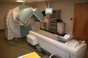 The UAB Division of Molecular Imaging and Therapeutics is equipped with a state-of-the-art GE Discovery LS PET-CT system, which will enable the resident to obtain experience with positron emission tomography in the clinical areas of cardiac, tumor, and brain. Another PET/CT system has been approved for the coming budget year, and as mentioned the VA Medical Center has recently added a PET/CT system as well. The primary PET radiopharmaceutical routinely used is 18F-FDG for the evaluation of regional glucose metabolism. Long term UAB plans include acquisition of a scintimammography camera, a SPECT/CT camera, and an on-site cyclotron as part of a research laboratory.
Application Process
We are accepting applications for Academic Year 2018 and 2019.
Requirements
Eligible applicants for a Molecular Imaging and Therapeutics fellowship position must complete (at a minimum) a PGY-1 clinical year in addition to an ACGME (or AOA, RCPSC, PCPQ) accredited residency. The fellow must pass USMLE (or COMLEX) steps I and II prior to beginning the residency. The resident must demonstrate a valid ECFMG certification (if applicable). Foreign citizens must be able to obtain a valid VISA, which UAB does sponsor, by the beginning of the fellowship contract.
Applications
If a resident wishes to be considered for an interview, the applicant will submit a UAB Application to our Program Coordinator. The above document also lists the minimum additional documents needed to complete the application (e.g., letters of recommendation, ECFMG certificates, etc). Keep in mind that we typically interview in the fall for positions open in the following summer. We will occasionally interview a year in advance on a limited basis.
Interview and Fellow Selection
Once all application materials are received, the Resident and Fellow Selection Committee will review the documents and determine which applicants will be granted an interview. This is based on a combination of the following: transcript reports (grades, test scores), written communication skills (for example, the applicant's personal statement), post-graduate experiences, research experience, clinical experience, imaging experience, certification by a subspecialty board, advanced degrees, recommendation letters, and Dean's letters. Only a limited number of applicants will be interviewed (approximately 4 to 6 per available position). Interviews will be scheduled directly with the applicant, and every attempt will be made to find a time convenient for the applicant. UAB is not able to reimburse applicants for their travel expenses. Once granted, the interview will consist of sequential meetings with numerous faculty members and a tour of the department. Time is also set aside for lunch with the current Nuclear Medicine residents and fellows, so the applicant will have an opportunity to get a resident and fellow's perspective of the program, and ask questions they may be too intimidated to ask a faculty interviewer. The interview day typically lasts from 8:30 AM to 2:30 PM. Following the interview, the Resident and Fellow Selection Committee makes a recommendation to the nuclear medicine faculty. The faculty will then come to a consensus as to who will be chosen.
The UAB Molecular Imaging and Therapeutics fellowship accepts residents and fellows outside of the national match. Once an offer is made and accepted, the fellow will receive a letter of intent. Contracts from the University will be sent as soon as they are available, usually in late spring, but is required to be at the UAB General Medical Education (GME) Office by July 1 or the first day of work. Residency positions are set for an academic year running July 1- June 30 of the following calendar year. We will accept off-cycle residents on a case-by-case basis, and as position availability and funding will allow.
General information can also be found at the following links:
UAB Diagnostic Radiology Department
University of Alabama at Birmingham
Alabama State Board of Medical Examiners
Leadership