The following patient was seen in the Emergency Department of Cayetano Heredia Hospital in Lima by the 2023 Gorgas Advanced Course participants.
History: A 77-year-old female patient presents to the Emergency Department with a 1-day history of frontal headache, which is followed by epistaxis of approximately 30cc in volume, which stops after applying pressure. On the day of admission, the headache persists, and the epistaxis recurs, but she is unable to stop the bleeding. She denies any trauma to the area. No weight loss or fever.
Epidemiology: Born and lives in Lima, no recent travel history. Works selling fruits in a market.
Past medical history: Hypertension, diagnosed 22 years ago, in irregular treatment with Losartan 50mg bid, and severe lower extremity venous insufficiency without treatment.
Physical Examination: BP 180/60 mmHg, HR 68 bpm, RR 16 rpm, Sat 98% on room air. Mucous membranes: No lesions in oral mucosa. Nasal examination findings are shown in Video A. Rest non-contributory.
UPCH Case Editors: Carlos Seas, Course Director / Paloma Carcamo, Associate Coordinator UAB Case Editors: David O. Freedman, Course Director Emeritus / German Henostroza, Course Director
{slide=CLICK HERE FOR DIAGNOSIS & DISCUSSION OF THE CASE ABOVE}
Diagnosis: Myiasis due to Cochliomyia hominivorax
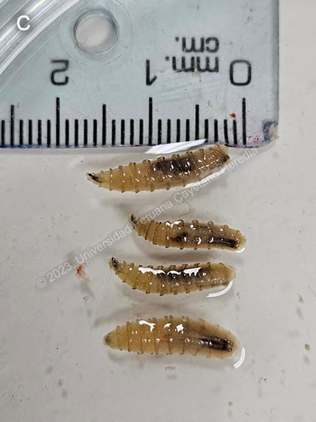
Discussion: Approximately thirty larvae were extracted from the patient’s nose (Video B). They were identified as Cochliomyia hominivorax larvae due to their smooth external aspect with dorsal tracheal trunks, and pigmented bands, as well as the posterior spiracles (Image C).
Myiasis refers to the infestation of vertebrates with the larval stages of flies of several different genera. Screwworm myiasis, caused by C. hominivorax in the New World and Chrysomya bezziana in the Old World, is associated with pre-existing wounds or with infestation of the mucous membranes. A recent report from Northern Peru documented that 5 of every 9 cases of myiasis in the region were due to C. hominivorax, and 4 of these 5 had associated cancers1. Another report from Ecuador documented 39 cases of myiasis, wherein 15 were caused by C. hominivorax2.
Furuncular myiasis is a different clinical entity, caused by larvae of Dermatobia hominis, the human bot fly, which is also present in the New World. The larvae of this species penetrate intact skin up to the subcutaneous tissue, forming an expanding boil-like lesion (see Case of the Week 2023-02). Cordylobia anthropophaga, known as the tumbu fly, can also cause furuncular myiasis but is endemic to tropical regions of Africa. Other genera of flies that can occasionally cause myiasis in humans include Cuterebra, Oestrus, and Wohlfhartia.
Screwworm myiasis can occur in any type of wound, including arthropod bites or wounds from diseases, or any mucous membrane, including nostrils, eye orbits, ears, mouth, genitalia, or the navels of newborns. Female flies of C. hominivorax lay eggs on the skin around wounds, which hatch 12-24 hours later and start to feed. The larvae develop for 4-10 days in the tissue up to a length of approximately 17mm, after which time they fall out and transform to pupa in the environment, where they will mature. An infested wound will attract gravid flies to lay more eggs, thus perpetuating the cycle3. The entire cycle usually takes 3 months but can take longer in cooler climates such as the Andes. Infested wounds typically have a distinctive odor, and may discharge blood, serum, or pus. Lesions in humans usually have a depth of 5 cm or more. It is important to promptly identify infestations because the screwworm can travel through the tissue beyond the subdermal layer, and perpetuation of the infection cycle can lead to massive infestation. There are no applicable serological tests, nor are they indicated in the identification of this disease. The diagnosis of myiasis is made by identification of the species of the extracted larva. However, in the case of this patient, further studies to assess for a predisposing underlying condition such as malignancy are warranted.
Treatment involves removal of the larvae and irrigation and debridement of affected tissue. The larvae have spines that they use as anchors, which may complicate extraction; local or general anesthesia may be required to ensure removal of all the flies4. Treatment with ivermectin, topically or orally, is safe and has been reported to be effective eradicating larvae as an adjuvant to manual extraction2,5. Ivermectin paralyzes the maggots and prevents further tissue damage, limiting the need for more extensive surgical debridement6.
Programs in the 1970s and 1980s aimed to eradicate New World screwworm in the United States through the use of sterile male flies, but outbreaks in deer have been reported as recently as 2017 in Florida, requiring new large interventions7. Similar campaigns are ongoing in Mexico and Central America.
At our institute, we see about ten outpatients per year with screwworm infestation; over the last ten years, ten patients have required hospitalization. Our patient was hospitalized and received treatment with ivermectin 200 ug/kg/day for two days after surgical removal of the larvae, as well as antibiotic coverage with clindamycin for bacterial superinfection. She continues to be monitored.
References 1. Failoc-Rojas VE, Molina-Ayasta C, Salazar-Zuloeta J, Samamé A, Silva-Díaz H. Case Report: Myiasis due to Cochliomyia hominivorax and Dermatobia hominis: Clinical and Pathological Differences between Two Species in Northern Peru. Am J Trop Med Hyg. 2018;98(1):150-153. doi:10.4269/ajtmh.16-0437 2. Calvopina M, Ortiz-Prado E, Castañeda B, Cueva I, Rodriguez-Hidalgo R, Cooper PJ. Human myiasis in Ecuador. PLOS Neglected Tropical Diseases. 2020;14(2):e0007858. doi:10.1371/journal.pntd.0007858 3. Bautista-Garfias CR, Aguilar-Marcelino L, Nogueda-Torres B. Myiasis infections in animals and men. Unique Scientific Publishers. 2023;3:20-27. doi:10.47278/book.oht/2023.72 4. Robbins K, Khachemoune A. Cutaneous myiasis: a review of the common types of myiasis. International Journal of Dermatology. 2010;49(10):1092-1098. doi:10.1111/j.1365-4632.2010.04577.x 5. Gealh WC, Ferreira GM, Farah GJ, Teodoro U, Camarini ET. Treatment of oral myiasis caused by Cochliomyia hominivorax: two cases treated with ivermectin. Br J Oral Maxillofac Surg. 2009;47(1):23-26. doi:10.1016/j.bjoms.2008.04.009 6. Osorio J, Moncada L, Molano A, Valderrama S, Gualtero S, Franco-Paredes C. Role of Ivermectin in the Treatment of Severe Orbital Myiasis Due to Cochliomyia hominivorax. Clinical Infectious Diseases. 2006;43(6):e57-e59. doi:10.1086/507038 7. Skoda SR, Phillips PL, Welch JB. Screwworm (Diptera: Calliphoridae) in the United States: Response to and Elimination of the 2016–2017 Outbreak in Florida. Journal of Medical Entomology. 2018;55(4):777-786. doi:10.1093/jme/tjy049