Imagine driving along I-20 west headed to Tuscaloosa for an Alabama football game.
Suddenly, a car slams on the brakes in front of you and you can’t avoid it. You crash into it. Both you and the other driver survive the crash and are taken to the hospital with broken bones, scrapes and bruises.
After the accident, you have normal feelings of fear, anxiety and anger, but over time, these feelings dissipate and you are left with a healthy respect for the dangers of the road. You are looking forward to a better drive to the next Alabama football game.
But, the other driver has a very different recovery experience. Their initial fears don’t go away, but instead persist and even escalate. They have flashbacks about the crash, nightmares that disrupt their sleep and they are too fearful to drive.
 These are symptoms of post-traumatic stress disorder, or PTSD. Why do two people who experience the same event have widely different responses?
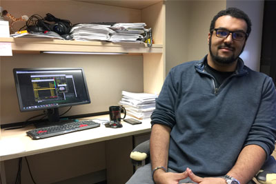
UAB doctoral student Nathaniel Harnett believes the answer to this question may lie in their brain.
The structure and function of the human brain has fascinated Harnett since his introduction to neuroscience in an undergraduate psychology class.
“The technology available today for scanning the brain is amazing,” said Harnett, a fifth-year graduate student in the UAB Department of Psychology’s Behavioral Neuroscience program. “We can look into the brain to learn why people have unique responses to events in their lives.”
In particular, Harnett is interested in how people respond to stressful or traumatic events, and why most individuals cope and recover, but some develop PTSD, which affects about 8 percent of U.S. citizens.
Harnett and his PhD mentor, Dr. David C. Knight, are using brain scans to identify people at risk for PTSD after they experience a traumatic event. Currently, treatments for PTSD are limited by gaps in our knowledge of the underlying neural mechanisms that lead to PTSD symptoms.
A diagnosis of PTSD is made by a psychiatrist or psychologist based on symptoms like re-experiencing the traumatic event, avoidance and arousal lasting longer than a few months that interfere with relationships, work and other aspects of daily life.
Once PTSD develops it can be hard to treat and involves counseling and medications.
“If we could identify individuals in danger of developing PTSD using a brain scan, we could potentially intervene to reduce or prevent symptoms of PTSD before they appear,” Harnett said.
Harnett’s research uses magnetic resonance imaging, or MRI, to look at brain structure and function. The brains of individuals with PTSD look different than those of individuals without PTSD.
Brain regions of interest are those involved in the normal expression of emotions and memory like the amygdala, hippocampus and prefrontal cortex. In order to identify differences in these brain regions after a traumatic event, Harnett and his colleagues recruited individuals who have recently experienced a traumatic injury (< 1 month ) from the UAB hospital trauma unit. They did not include people who have any other brain injury or condition that could affect their mental function.
 They conducted brain scans with research participants that look at brain structure and function and assess PTSD symptoms with a self-report measure called the Post-traumatic Diagnostic Scale (PDS). Study participants reported their PTSD symptoms with the self-report tool again at three months and six months after they begin the study.
Harnett and his colleagues are trying to find a “signature of PTSD” or PTSD susceptibility by using this multimodal imaging approach that includes brain structure, function and biochemistry.
“We are discovering that differences in brain structure, function and biochemistry early after trauma are related to the development of PTSD,” Harnett said.
For example, decreases in size of the hippocampus could make it hard to discriminate between past and present experiences; decreases in prefrontal cortex size could impair fear-regulating processes; and hyperactivity in the amygdala could lead to anxiety. More work is necessary to better understand these changes and whether there are relationships between them.
Harnett emphasized that the most rewarding aspects of this research include working with an under-researched population, and the prospect of finding ways to reduce disability associated with PTSD and improve quality of life for affected individuals.
Ultimately, Harnett and his colleagues envision rehabilitation strategies that could be used before PTSD develops with individuals at risk based on their brain scans. Techniques like transcranial magnetic stimulation (a method of externally stimulating the brain) may be useful to positively influence communication networks in the brain and reduce PTSD symptoms.
Harnett plans to continue this line of research during his postdoctoral research fellowship working under Kerry Ressler at McLean Hospital in Boston. His long-term goal is to be a faculty member and the principal investigator of his own lab. He wants to train and inspire the next generation of scientists.
“This is a very cool area of science,” Harnett said. “How many different fields out there get to look at your brain and also have the potential to change lives?”